 Movie
Movie Controller

[English] 日本語
 Yorodumi
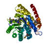
- PDB-3brc: Crystal structure of a conserved protein of unknown function from... -
+ Open data

- Basic information
| Entry | Database: PDB / ID: 3brc | ||||||
|---|---|---|---|---|---|---|---|
| Title | Crystal structure of a conserved protein of unknown function from Methanobacterium thermoautotrophicum | ||||||
 Components Components | Conserved protein of unknown function | ||||||
 Keywords Keywords | STRUCTURAL GENOMICS / UNKNOWN FUNCTION / Conserved protein / Methanobacterium thermoautotrophicum / MCSG / PSI-2 / Protein Structure Initiative / Midwest Center for Structural Genomics | ||||||
| Function / homology |  Function and homology information Function and homology informationFeGP cofactor biosynthesis protein HcgB, guanylyltransferase / FeGP cofactor biosynthesis protein HcgB, guanylyltransferase / B12-dependent dehydatase associated subunit / Helix hairpin bin / B12-dependent dehydratases, beta subunit / PUA domain profile. / Helix Hairpins / Rossmann fold / Orthogonal Bundle / 3-Layer(aba) Sandwich ...FeGP cofactor biosynthesis protein HcgB, guanylyltransferase / FeGP cofactor biosynthesis protein HcgB, guanylyltransferase / B12-dependent dehydatase associated subunit / Helix hairpin bin / B12-dependent dehydratases, beta subunit / PUA domain profile. / Helix Hairpins / Rossmann fold / Orthogonal Bundle / 3-Layer(aba) Sandwich / Mainly Alpha / Alpha Beta Similarity search - Domain/homology | ||||||
| Biological species |   Methanothermobacter thermautotrophicus (archaea) Methanothermobacter thermautotrophicus (archaea) | ||||||
| Method |  X-RAY DIFFRACTION / SYNCHROTRON / SAD / Resolution: 1.6 Å X-RAY DIFFRACTION / SYNCHROTRON / SAD / Resolution: 1.6 Å | ||||||
 Authors Authors | Zhang, R. / Evdokimova, E. / Kudritska, M. / Savchenko, A. / Edwards, A.M. / Joachimiak, A. / Midwest Center for Structural Genomics (MCSG) | ||||||
 Citation Citation | Journal: To be Published Title: The crystal structure of a conserved protein of unknown function from Methanobacterium thermoautotrophicum. Authors: Zhang, R. / Evdokimova, E. / Kudritska, M. / Savchenko, A. / Edwards, A.M. / Joachimiak, A. | ||||||
| History |
|
- Structure visualization
| Structure viewer | Molecule: MolmilJmol/JSmol |
|---|
- Downloads & links
-Download
| PDBx/mmCIF format | 3brc.cif.gz | 139.4 KB | Display | PDBx/mmCIF format |
|---|---|---|---|---|
| PDB format | pdb3brc.ent.gz | 110.4 KB | Display | PDB format |
| PDBx/mmJSON format | 3brc.json.gz | Tree view | PDBx/mmJSON format | |
| Others |  Other downloads Other downloads |
-Validation report
| Arichive directory | https://data.pdbj.org/pub/pdb/validation_reports/br/3brcftp://data.pdbj.org/pub/pdb/validation_reports/br/3brc | HTTPS FTP |
|---|
-Related structure data
| Similar structure data | |
|---|---|
| Other databases |


-Links
 PDBj
PDBj- Assembly
| Deposited unit | 
| ||||||||
|---|---|---|---|---|---|---|---|---|---|
| 1 |
| ||||||||
| 2 | 
| ||||||||
| Unit cell |
| ||||||||
| Components on special symmetry positions |
|
-Components
| #1: Protein | Mass: 17893.459 Da / Num. of mol.: 2 Source method: isolated from a genetically manipulated source Source: (gene. exp.) Methanothermobacter thermautotrophicus (archaea)Strain: Delta H / Gene: MTH1144, GI:2622249 / Plasmid: pDM68 / Species (production host): Escherichia coli / Production host:  #2: Chemical |   Mass: 94.971 Da / Num. of mol.: 3 / Source method: obtained synthetically / Formula: PO4 Mass: 94.971 Da / Num. of mol.: 3 / Source method: obtained synthetically / Formula: PO4#3: Water | ChemComp-HOH / |  Mass: 18.015 Da / Num. of mol.: 226 / Source method: isolated from a natural source / Formula: H2O Mass: 18.015 Da / Num. of mol.: 226 / Source method: isolated from a natural source / Formula: H2OHas protein modification | Y | |
|---|
-Experimental details
-Experiment
| Experiment | Method: X-RAY DIFFRACTION / Number of used crystals: 1 |
|---|
- Sample preparation
| Crystal | Density Matthews: 2.05 Å3/Da / Density % sol: 39.98 % |
|---|---|
| Crystal grow | Temperature: 289 K / Method: vapor diffusion, sitting drop Details: 0.1M Ammonium phosphate, 16% PEG 3350, 2% Glycerol, VAPOR DIFFUSION, SITTING DROP, temperature 289K |
-Data collection
| Diffraction | Mean temperature: 100 K |
|---|---|
| Diffraction source | Source: SYNCHROTRON / Site: APS  / Beamline: 19-ID / Wavelength: 0.9794 Å / Beamline: 19-ID / Wavelength: 0.9794 Å |
| Detector | Type: ADSC QUANTUM 315 / Detector: CCD / Date: Jul 3, 2007 / Details: Mirrors |
| Radiation | Monochromator: Si 111 channel / Protocol: SINGLE WAVELENGTH / Monochromatic (M) / Laue (L): M / Scattering type: x-ray |
| Radiation wavelength | Wavelength: 0.9794 Å / Relative weight: 1 |
| Reflection | Resolution: 1.6→88.52 Å / Num. all: 36473 / Num. obs: 32286 / % possible obs: 88.52 % / Observed criterion σ(I): 2 / Redundancy: 4.4 % / Biso Wilson estimate: 18 Å2 / Rmerge(I) obs: 0.09 / Net I/σ(I): 27.05 |
| Reflection shell | Resolution: 1.6→1.642 Å / Redundancy: 2 % / Rmerge(I) obs: 0.378 / Mean I/σ(I) obs: 1.35 / Num. unique all: 2852 / % possible all: 40.92 |
- Processing
| Software |
| |||||||||||||||||||||||||||||||||||||||||||||||||||||||||||||||||||||||||||||||||||||||||||||||||||||||||||||||||||||||||||||
|---|---|---|---|---|---|---|---|---|---|---|---|---|---|---|---|---|---|---|---|---|---|---|---|---|---|---|---|---|---|---|---|---|---|---|---|---|---|---|---|---|---|---|---|---|---|---|---|---|---|---|---|---|---|---|---|---|---|---|---|---|---|---|---|---|---|---|---|---|---|---|---|---|---|---|---|---|---|---|---|---|---|---|---|---|---|---|---|---|---|---|---|---|---|---|---|---|---|---|---|---|---|---|---|---|---|---|---|---|---|---|---|---|---|---|---|---|---|---|---|---|---|---|---|---|---|---|
| Refinement | Method to determine structure: SAD / Resolution: 1.6→56.08 Å / Cor.coef. Fo:Fc: 0.956 / Cor.coef. Fo:Fc free: 0.931 / SU B: 4.177 / SU ML: 0.068 / Cross valid method: THROUGHOUT / σ(F): 0 / σ(I): 0 / ESU R: 0.178 / ESU R Free: 0.116 Stereochemistry target values: MAXIMUM LIKELIHOOD WITH PHASES Details: HYDROGENS HAVE BEEN ADDED IN THE RIDING POSITIONS
| |||||||||||||||||||||||||||||||||||||||||||||||||||||||||||||||||||||||||||||||||||||||||||||||||||||||||||||||||||||||||||||
| Solvent computation | Ion probe radii: 0.8 Å / Shrinkage radii: 0.8 Å / VDW probe radii: 1.2 Å / Solvent model: MASK | |||||||||||||||||||||||||||||||||||||||||||||||||||||||||||||||||||||||||||||||||||||||||||||||||||||||||||||||||||||||||||||
| Displacement parameters | Biso mean: 17.697 Å2
| |||||||||||||||||||||||||||||||||||||||||||||||||||||||||||||||||||||||||||||||||||||||||||||||||||||||||||||||||||||||||||||
| Refinement step | Cycle: LAST / Resolution: 1.6→56.08 Å
| |||||||||||||||||||||||||||||||||||||||||||||||||||||||||||||||||||||||||||||||||||||||||||||||||||||||||||||||||||||||||||||
| Refine LS restraints |
| |||||||||||||||||||||||||||||||||||||||||||||||||||||||||||||||||||||||||||||||||||||||||||||||||||||||||||||||||||||||||||||
| LS refinement shell | Resolution: 1.6→1.64 Å / Total num. of bins used: 20
| |||||||||||||||||||||||||||||||||||||||||||||||||||||||||||||||||||||||||||||||||||||||||||||||||||||||||||||||||||||||||||||
| Refinement TLS params. | Method: refined / Origin x: 6.0672 Å / Origin y: 1.8108 Å / Origin z: 12.2825 Å
| |||||||||||||||||||||||||||||||||||||||||||||||||||||||||||||||||||||||||||||||||||||||||||||||||||||||||||||||||||||||||||||
| Refinement TLS group |
|
