 Movie
Movie Controller

[English] 日本語
 Yorodumi
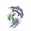
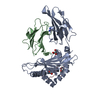
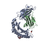
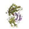
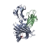
- PDB-3bp7: The high resolution crystal structure of HLA-B*2709 in complex wi... -
+ Open data

- Basic information
| Entry | Database: PDB / ID: 3bp7 | ||||||
|---|---|---|---|---|---|---|---|
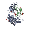
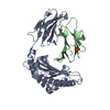
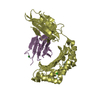
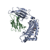
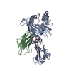
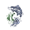
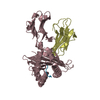
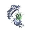
| Title | The high resolution crystal structure of HLA-B*2709 in complex with a Cathepsin A signal sequence peptide, pCatA | ||||||
 Components Components |
| ||||||
 Keywords Keywords | IMMUNE SYSTEM / Major Histocompatibility Complex / MHC / Human Leukocyte Antigen / HLA / HLA-B*2709 / HLA-B2709 / beta-2-microglobulin / b2m / Cathepsin A signal sequence / pCatA / Ankylosing Spondylitis / Glycoprotein / Host-virus interaction / Immune response / Membrane / MHC I / Transmembrane / Disease mutation / Glycation / Immunoglobulin domain / Pyrrolidone carboxylic acid / Secreted / Carboxypeptidase / Hydrolase / Lysosome / Protease / Zymogen | ||||||
| Function / homology |  Function and homology information Function and homology informationcarboxypeptidase C / Defective NEU1 causes sialidosis / serine-type carboxypeptidase activity / Sialic acid metabolism / regulation of chaperone-mediated autophagy / Glycosphingolipid catabolism / regulation of interleukin-12 production / regulation of dendritic cell differentiation / regulation of T cell anergy / regulation of interleukin-6 production ...carboxypeptidase C / Defective NEU1 causes sialidosis / serine-type carboxypeptidase activity / Sialic acid metabolism / regulation of chaperone-mediated autophagy / Glycosphingolipid catabolism / regulation of interleukin-12 production / regulation of dendritic cell differentiation / regulation of T cell anergy / regulation of interleukin-6 production / negative regulation of chaperone-mediated autophagy / protection from natural killer cell mediated cytotoxicity / TAP binding / detection of bacterium / antigen processing and presentation of endogenous peptide antigen via MHC class Ib / antigen processing and presentation of endogenous peptide antigen via MHC class I via ER pathway, TAP-independent / carboxypeptidase activity / MHC class II antigen presentation / lysosomal lumen / early endosome lumen / Nef mediated downregulation of MHC class I complex cell surface expression / DAP12 interactions / secretory granule membrane / T cell mediated cytotoxicity / Endosomal/Vacuolar pathway / Antigen Presentation: Folding, assembly and peptide loading of class I MHC / lumenal side of endoplasmic reticulum membrane / regulation of iron ion transport / cellular response to iron(III) ion / antigen processing and presentation of exogenous protein antigen via MHC class Ib, TAP-dependent / negative regulation of iron ion transport / negative regulation of forebrain neuron differentiation / regulation of erythrocyte differentiation / peptide antigen assembly with MHC class I protein complex / ER to Golgi transport vesicle membrane / response to molecule of bacterial origin / defense response / regulation of protein stability / HFE-transferrin receptor complex / MHC class I peptide loading complex / transferrin transport / negative regulation of receptor-mediated endocytosis / cellular response to iron ion / positive regulation of T cell cytokine production / antigen processing and presentation of endogenous peptide antigen via MHC class I / MHC class I protein complex / intracellular protein transport / peptide antigen assembly with MHC class II protein complex / negative regulation of neurogenesis / multicellular organismal-level iron ion homeostasis / cellular response to nicotine / MHC class II protein complex / positive regulation of receptor-mediated endocytosis / positive regulation of T cell mediated cytotoxicity / negative regulation of epithelial cell proliferation / specific granule lumen / antigen processing and presentation of exogenous peptide antigen via MHC class II / positive regulation of immune response / peptide antigen binding / phagocytic vesicle membrane / recycling endosome membrane / enzyme activator activity / positive regulation of T cell activation / Interferon gamma signaling / Immunoregulatory interactions between a Lymphoid and a non-Lymphoid cell / Interferon alpha/beta signaling / Modulation by Mtb of host immune system / azurophil granule lumen / sensory perception of smell / positive regulation of cellular senescence / tertiary granule lumen / MHC class II protein complex binding / T cell differentiation in thymus / DAP12 signaling / late endosome membrane / negative regulation of neuron projection development / protein refolding / protein-folding chaperone binding / ER-Phagosome pathway / early endosome membrane / amyloid fibril formation / protein homotetramerization / intracellular iron ion homeostasis / adaptive immune response / learning or memory / lysosome / immune response / endoplasmic reticulum lumen / Amyloid fiber formation / Golgi membrane / external side of plasma membrane / signaling receptor binding / innate immune response / lysosomal membrane / focal adhesion / Neutrophil degranulation / SARS-CoV-2 activates/modulates innate and adaptive immune responses / structural molecule activity / Golgi apparatus / cell surface Similarity search - Function | ||||||
| Biological species |  Homo sapiens (human)Homo Sapiens (human) Homo sapiens (human)Homo Sapiens (human) | ||||||
| Method |  X-RAY DIFFRACTION / SYNCHROTRON / MOLECULAR REPLACEMENT / molecular replacement / Resolution: 1.8 Å X-RAY DIFFRACTION / SYNCHROTRON / MOLECULAR REPLACEMENT / molecular replacement / Resolution: 1.8 Å | ||||||
 Authors Authors | Kumar, P. / Vahedi-Faridi, A. / Saenger, W. / Uchanska-Ziegler, B. / Ziegler, A. | ||||||
 Citation Citation | Journal: J.Biol.Chem. / Year: 2009 Title: Structural basis for T cell alloreactivity among three HLA-B14 and HLA-B27 antigens Authors: Kumar, P. / Vahedi-Faridi, A. / Saenger, W. / Merino, E. / Lopez de Castro, J.A. / Uchanska-Ziegler, B. / Ziegler, A. | ||||||
| History |
|
- Structure visualization
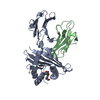
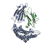
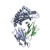
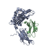
| Structure viewer | Molecule: MolmilJmol/JSmol |
|---|
- Downloads & links
-Download
| PDBx/mmCIF format | 3bp7.cif.gz | 106.2 KB | Display | PDBx/mmCIF format |
|---|---|---|---|---|
| PDB format | pdb3bp7.ent.gz | 78.9 KB | Display | PDB format |
| PDBx/mmJSON format | 3bp7.json.gz | Tree view | PDBx/mmJSON format | |
| Others |  Other downloads Other downloads |
-Validation report
| Arichive directory | https://data.pdbj.org/pub/pdb/validation_reports/bp/3bp7ftp://data.pdbj.org/pub/pdb/validation_reports/bp/3bp7 | HTTPS FTP |
|---|
-Related structure data
| Related structure data | 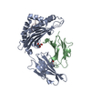 3bp4C 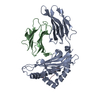 3bvnC  3bxnC 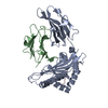 1jgeS C: citing same article ( S: Starting model for refinement |
|---|---|
| Similar structure data |


-Links
 PDBj
PDBj


- Assembly
| Deposited unit | 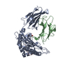
| ||||||||
|---|---|---|---|---|---|---|---|---|---|
| 1 |
| ||||||||
| Unit cell |
|
-Components
| #1: Protein | Mass: 31951.219 Da / Num. of mol.: 1 Fragment: HLA-B*2709 extracellular domain, UNP residues 25-300 Source method: isolated from a genetically manipulated source Source: (gene. exp.) Homo sapiens (human) / Plasmid: pHN1 / Production host:  |
|---|---|
| #2: Protein | Mass: 11879.356 Da / Num. of mol.: 1 Source method: isolated from a genetically manipulated source Source: (gene. exp.) Homo sapiens (human) / Plasmid: pHN1 / Production host: |
| #3: Protein/peptide | Mass: 982.198 Da / Num. of mol.: 1 / Fragment: Cathepsin A signal sequence, UNP residues 2-10 / Source method: obtained synthetically Details: Cathepsin A signal sequence peptide, pCatA, chemically synthesized, This sequence occurs naturally in humans. Source: (synth.) Homo Sapiens (human) / References: UniProt: P10619 |
| #4: Chemical | ChemComp-GOL /   Mass: 92.094 Da / Num. of mol.: 1 / Source method: obtained synthetically / Formula: C3H8O3 Mass: 92.094 Da / Num. of mol.: 1 / Source method: obtained synthetically / Formula: C3H8O3 |
| #5: Water | ChemComp-HOH /  Mass: 18.015 Da / Num. of mol.: 513 / Source method: isolated from a natural source / Formula: H2O Mass: 18.015 Da / Num. of mol.: 513 / Source method: isolated from a natural source / Formula: H2O |
| Has protein modification | Y |
| Sequence details | THE 140TH RESIDUE IS HIS IN ALLELE B*2709. |
-Experimental details
-Experiment
| Experiment | Method: X-RAY DIFFRACTION / Number of used crystals: 1 |
|---|
- Sample preparation
| Crystal | Density Matthews: 2.63 Å3/Da / Density % sol: 53.16 % |
|---|---|
| Crystal grow | Temperature: 291 K / Method: vapor diffusion, hanging drop / pH: 7.5 Details: 23.5% PEG 8000, 0.1M Tris buffer pH 7.5, VAPOR DIFFUSION, HANGING DROP, temperature 291K |
-Data collection
| Diffraction | Mean temperature: 100 K |
|---|---|
| Diffraction source | Source: SYNCHROTRON / Site: BESSY  / Beamline: 14.2 / Wavelength: 0.95373 Å / Beamline: 14.2 / Wavelength: 0.95373 Å |
| Detector | Type: MARRESEARCH / Detector: CCD / Date: Jan 18, 2006 / Details: Mirrors |
| Radiation | Monochromator: Si-111 crystal / Protocol: SINGLE WAVELENGTH / Monochromatic (M) / Laue (L): M / Scattering type: x-ray |
| Radiation wavelength | Wavelength: 0.95373 Å / Relative weight: 1 |
| Reflection | Resolution: 1.79→50 Å / Num. obs: 41600 / % possible obs: 93.3 % / Redundancy: 3.6 % / Biso Wilson estimate: 19.9 Å2 / Rmerge(I) obs: 0.057 / Χ2: 0.796 / Net I/σ(I): 19.4 |
| Reflection shell | Resolution: 1.79→1.85 Å / Redundancy: 3.3 % / Rmerge(I) obs: 0.331 / Mean I/σ(I) obs: 3.07 / Num. unique all: 3694 / Χ2: 0.684 / % possible all: 84.3 |
-Phasing
| Phasing | Method: molecular replacement | |||||||||
|---|---|---|---|---|---|---|---|---|---|---|
| Phasing MR | Rfactor: 0.47 / Cor.coef. Fo:Fc: 0.463
|
- Processing
| Software |
| ||||||||||||||||||||||||||||||||||||||||||||||||||||||||||||||||||||||||||||||||||||||||||||||||||||||||||||||||||||||||
|---|---|---|---|---|---|---|---|---|---|---|---|---|---|---|---|---|---|---|---|---|---|---|---|---|---|---|---|---|---|---|---|---|---|---|---|---|---|---|---|---|---|---|---|---|---|---|---|---|---|---|---|---|---|---|---|---|---|---|---|---|---|---|---|---|---|---|---|---|---|---|---|---|---|---|---|---|---|---|---|---|---|---|---|---|---|---|---|---|---|---|---|---|---|---|---|---|---|---|---|---|---|---|---|---|---|---|---|---|---|---|---|---|---|---|---|---|---|---|---|---|---|
| Refinement | Method to determine structure: MOLECULAR REPLACEMENT Starting model: PDB ENTRY 1JGE Resolution: 1.8→46.08 Å / Cor.coef. Fo:Fc: 0.956 / Cor.coef. Fo:Fc free: 0.937 / SU B: 2.24 / SU ML: 0.071 / Cross valid method: THROUGHOUT / ESU R: 0.121 / ESU R Free: 0.117 / Stereochemistry target values: MAXIMUM LIKELIHOOD / Details: HYDROGENS HAVE BEEN ADDED IN THE RIDING POSITIONS
| ||||||||||||||||||||||||||||||||||||||||||||||||||||||||||||||||||||||||||||||||||||||||||||||||||||||||||||||||||||||||
| Solvent computation | Ion probe radii: 0.8 Å / Shrinkage radii: 0.8 Å / VDW probe radii: 1.2 Å / Solvent model: BABINET MODEL WITH MASK | ||||||||||||||||||||||||||||||||||||||||||||||||||||||||||||||||||||||||||||||||||||||||||||||||||||||||||||||||||||||||
| Displacement parameters | Biso mean: 14.028 Å2
| ||||||||||||||||||||||||||||||||||||||||||||||||||||||||||||||||||||||||||||||||||||||||||||||||||||||||||||||||||||||||
| Refinement step | Cycle: LAST / Resolution: 1.8→46.08 Å
| ||||||||||||||||||||||||||||||||||||||||||||||||||||||||||||||||||||||||||||||||||||||||||||||||||||||||||||||||||||||||
| Refine LS restraints |
| ||||||||||||||||||||||||||||||||||||||||||||||||||||||||||||||||||||||||||||||||||||||||||||||||||||||||||||||||||||||||
| LS refinement shell | Resolution: 1.797→1.843 Å / Total num. of bins used: 20
|
