 Movie
Movie Controller


+ Open data

- Basic information
| Entry | Database: PDB / ID: 3bm3 | ||||||
|---|---|---|---|---|---|---|---|
| Title | Restriction endonuclease PspGI-substrate DNA complex | ||||||
 Components Components |
| ||||||
 Keywords Keywords | HYDROLASE/DNA / ENDONUCLEASE-DNA COMPLEX / RESTRICTION ENZYME / PSPGI / BASE FLIPPING / HYDROLASE-DNA COMPLEX | ||||||
| Function / homology |  Function and homology information Function and homology informationtype II site-specific deoxyribonuclease activity / DNA restriction-modification system / DNA binding Similarity search - Function | ||||||
| Biological species |   Pyrococcus sp. GI-H (archaea) Pyrococcus sp. GI-H (archaea) | ||||||
| Method |  X-RAY DIFFRACTION / SYNCHROTRON / Resolution: 1.7 Å X-RAY DIFFRACTION / SYNCHROTRON / Resolution: 1.7 Å | ||||||
 Authors Authors | Szczepanowski, R.H. / Carpenter, M. / Czapinska, H. / Tamulaitis, G. / Siksnys, V. / Bhagwat, A. / Bochtler, M. | ||||||
 Citation Citation | Journal: To be Published Title: A direct crystallographic demonstration that Type II restriction endonuclease PspGI flips nucleotides Authors: Szczepanowski, R.H. / Carpenter, M. / Czapinska, H. / Tamulaitis, G. / Siksnys, V. / Bhagwat, A. / Bochtler, M. #1: Journal: Nucleic Acids Res. / Year: 2007 Title: Nucleotide flipping by restriction enzymes analyzed by 2-aminopurine steady-state fluorescence. Authors: Tamulaitis, G. / Zaremba, M. / Szczepanowski, R.H. / Bochtler, M. / Siksnys, V. #2: Journal: Nucleic Acids Res. / Year: 2006 Title: Sequence-dependent enhancement of hydrolytic deamination of cytosines in DNA by the restriction enzyme PspGI. Authors: Carpenter, M. / Divvela, P. / Pingoud, V. / Bujnicki, J. / Bhagwat, A.S. #3: Journal: J.Mol.Biol. / Year: 2003 Title: PspGI, a type II restriction endonuclease from the extreme thermophile Pyrococcus sp.: structural and functional studies to investigate an evolutionary relationship with several mesophilic restriction enzymes. Authors: Pingoud, V. / Conzelmann, C. / Kinzebach, S. / Sudina, A. / Metelev, V. / Kubareva, E. / Bujnicki, J.M. / Lurz, R. / Luder, G. / Xu, S.Y. / Pingoud, A. #4: Journal: Appl.Environ.Microbiol. / Year: 1998 Title: Characterization of an extremely thermostable restriction enzyme, PspGI, from a Pyrococcus strain and cloning of the PspGI restriction-modification system in Escherichia coli. Authors: Morgan, R. / Xiao, J. / Xu, S. | ||||||
| History |
|
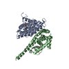
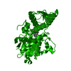
- Structure visualization
| Structure viewer | Molecule: MolmilJmol/JSmol |
|---|
- Downloads & links
-Download
| PDBx/mmCIF format | 3bm3.cif.gz | 143.2 KB | Display | PDBx/mmCIF format |
|---|---|---|---|---|
| PDB format | pdb3bm3.ent.gz | 111.3 KB | Display | PDB format |
| PDBx/mmJSON format | 3bm3.json.gz | Tree view | PDBx/mmJSON format | |
| Others |  Other downloads Other downloads |
-Validation report
| Summary document | 3bm3_validation.pdf.gz | 469.6 KB | Display | wwPDB validaton report |
|---|---|---|---|---|
| Full document | 3bm3_full_validation.pdf.gz | 476.7 KB | Display | |
| Data in XML | 3bm3_validation.xml.gz | 24.1 KB | Display | |
| Data in CIF | 3bm3_validation.cif.gz | 35.3 KB | Display | |
| Arichive directory | https://data.pdbj.org/pub/pdb/validation_reports/bm/3bm3ftp://data.pdbj.org/pub/pdb/validation_reports/bm/3bm3 | HTTPS FTP |
-Related structure data
| Similar structure data |
|---|

-Links
 PDBj
PDBj


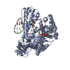
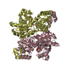
- Assembly
| Deposited unit | 
| ||||||||
|---|---|---|---|---|---|---|---|---|---|
| 1 |
| ||||||||
| Unit cell |
| ||||||||
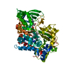
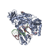
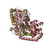
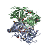
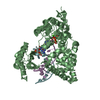
| Details | THE TETRAMER (ACCORDING TO PDB CONVENTIONS) IS A COMPLEX OF THE DIMERIC RESTRICTION ENZYME WITH ITS SUBSTRATE, DOUBLE STRANDED DNA. |
-Components
| #1: DNA chain | Mass: 3318.187 Da / Num. of mol.: 1 / Source method: obtained synthetically | ||||||
|---|---|---|---|---|---|---|---|
| #2: DNA chain | Mass: 3389.221 Da / Num. of mol.: 1 / Source method: obtained synthetically | ||||||
| #3: Protein | Mass: 32215.279 Da / Num. of mol.: 2 / Mutation: D138A, A146T Source method: isolated from a genetically manipulated source Source: (gene. exp.) Pyrococcus sp. GI-H (archaea) / Plasmid: pET21a / Production host:  #4: Chemical | ChemComp-CIT / |   Mass: 192.124 Da / Num. of mol.: 1 / Source method: obtained synthetically / Formula: C6H8O7 Mass: 192.124 Da / Num. of mol.: 1 / Source method: obtained synthetically / Formula: C6H8O7#5: Water | ChemComp-HOH / |  Mass: 18.015 Da / Num. of mol.: 293 / Source method: isolated from a natural source / Formula: H2O Mass: 18.015 Da / Num. of mol.: 293 / Source method: isolated from a natural source / Formula: H2OHas protein modification | Y | |
-Experimental details
-Experiment
| Experiment | Method: X-RAY DIFFRACTION / Number of used crystals: 1 |
|---|
- Sample preparation
| Crystal | Density Matthews: 2.01 Å3/Da / Density % sol: 38.84 % | ||||||||||||||||||||||||||||
|---|---|---|---|---|---|---|---|---|---|---|---|---|---|---|---|---|---|---|---|---|---|---|---|---|---|---|---|---|---|
| Crystal grow | Temperature: 294 K / Method: vapor diffusion, sitting drop / pH: 4 Details: 20% MPD, 0.1 M citric acid, 0.1 M cobalt (II) chloride as an additive, pH 4.0, VAPOR DIFFUSION, SITTING DROP, temperature 294K | ||||||||||||||||||||||||||||
| Components of the solutions |
|
-Data collection
| Diffraction | Mean temperature: 100 K |
|---|---|
| Diffraction source | Source: SYNCHROTRON / Site: MPG/DESY, HAMBURG  / Beamline: BW6 / Wavelength: 0.976 Å / Beamline: BW6 / Wavelength: 0.976 Å |
| Detector | Type: MARRESEARCH / Detector: CCD / Date: Aug 16, 2007 / Details: BENT MIRROR |
| Radiation | Monochromator: TRIANGULAR MONOCHROMATOR / Protocol: SINGLE WAVELENGTH / Monochromatic (M) / Laue (L): M / Scattering type: x-ray |
| Radiation wavelength | Wavelength: 0.976 Å / Relative weight: 1 |
| Reflection | Resolution: 1.7→20 Å / Num. all: 63138 / Num. obs: 63138 / % possible obs: 98.7 % / Redundancy: 3.3 % / Biso Wilson estimate: 20.7 Å2 / Rsym value: 0.065 / Net I/σ(I): 21.3 |
| Reflection shell | Resolution: 1.7→1.72 Å / Redundancy: 3.1 % / Mean I/σ(I) obs: 3.2 / Num. unique all: 2460 / Rsym value: 0.254 / % possible all: 98 |
- Processing
| Software |
| ||||||||||||||||||||||||||||||||||||||||||||||||||||||||||||||||||||||||||||||||||||||||||||||||||||||||||||||||||||||||||||||||||||||||||||||||||||||||||||||||||||||||||
|---|---|---|---|---|---|---|---|---|---|---|---|---|---|---|---|---|---|---|---|---|---|---|---|---|---|---|---|---|---|---|---|---|---|---|---|---|---|---|---|---|---|---|---|---|---|---|---|---|---|---|---|---|---|---|---|---|---|---|---|---|---|---|---|---|---|---|---|---|---|---|---|---|---|---|---|---|---|---|---|---|---|---|---|---|---|---|---|---|---|---|---|---|---|---|---|---|---|---|---|---|---|---|---|---|---|---|---|---|---|---|---|---|---|---|---|---|---|---|---|---|---|---|---|---|---|---|---|---|---|---|---|---|---|---|---|---|---|---|---|---|---|---|---|---|---|---|---|---|---|---|---|---|---|---|---|---|---|---|---|---|---|---|---|---|---|---|---|---|---|---|---|
| Refinement | Resolution: 1.7→19.38 Å / Cor.coef. Fo:Fc: 0.962 / Cor.coef. Fo:Fc free: 0.955 / TLS residual ADP flag: LIKELY RESIDUAL / Cross valid method: THROUGHOUT / ESU R: 0.109 / ESU R Free: 0.099 / Stereochemistry target values: MAXIMUM LIKELIHOOD Details: CNS HAS BEEN USED FOR DNA REFINEMENT. NO CONSTRAINTS FOR SUGAR PUCKER WERE APPLIED. RESIDUES AT THE ENDS OF THE DNA DUPLEX ( 5 AND 5 IN CHAIN C AND -6 AND -5 IN CHAIN D) HAVE POOR DENSITY. ...Details: CNS HAS BEEN USED FOR DNA REFINEMENT. NO CONSTRAINTS FOR SUGAR PUCKER WERE APPLIED. RESIDUES AT THE ENDS OF THE DNA DUPLEX ( 5 AND 5 IN CHAIN C AND -6 AND -5 IN CHAIN D) HAVE POOR DENSITY. HYDROGENS HAVE BEEN ADDED IN THE RIDING POSITIONS. TLS REFINEMENT HAS BEEN USED.
| ||||||||||||||||||||||||||||||||||||||||||||||||||||||||||||||||||||||||||||||||||||||||||||||||||||||||||||||||||||||||||||||||||||||||||||||||||||||||||||||||||||||||||
| Solvent computation | Ion probe radii: 0.8 Å / Shrinkage radii: 0.8 Å / VDW probe radii: 1.4 Å / Solvent model: MASK | ||||||||||||||||||||||||||||||||||||||||||||||||||||||||||||||||||||||||||||||||||||||||||||||||||||||||||||||||||||||||||||||||||||||||||||||||||||||||||||||||||||||||||
| Displacement parameters | Biso mean: 17.9 Å2
| ||||||||||||||||||||||||||||||||||||||||||||||||||||||||||||||||||||||||||||||||||||||||||||||||||||||||||||||||||||||||||||||||||||||||||||||||||||||||||||||||||||||||||
| Refinement step | Cycle: LAST / Resolution: 1.7→19.38 Å
| ||||||||||||||||||||||||||||||||||||||||||||||||||||||||||||||||||||||||||||||||||||||||||||||||||||||||||||||||||||||||||||||||||||||||||||||||||||||||||||||||||||||||||
| Refine LS restraints |
| ||||||||||||||||||||||||||||||||||||||||||||||||||||||||||||||||||||||||||||||||||||||||||||||||||||||||||||||||||||||||||||||||||||||||||||||||||||||||||||||||||||||||||
| LS refinement shell | Resolution: 1.7→1.744 Å / Total num. of bins used: 20
| ||||||||||||||||||||||||||||||||||||||||||||||||||||||||||||||||||||||||||||||||||||||||||||||||||||||||||||||||||||||||||||||||||||||||||||||||||||||||||||||||||||||||||
| Refinement TLS params. | Method: refined / Refine-ID: X-RAY DIFFRACTION
| ||||||||||||||||||||||||||||||||||||||||||||||||||||||||||||||||||||||||||||||||||||||||||||||||||||||||||||||||||||||||||||||||||||||||||||||||||||||||||||||||||||||||||
| Refinement TLS group |
|
