- PDB-3b9t: Crystal structure of predicted acetamidase/formamidase (YP_546212... -
+
Open data
ID or keywords:
Loading...
-
Basic information
Entry
Database: PDB / ID: 3b9t
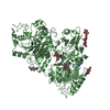
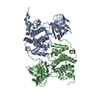
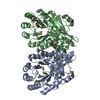
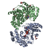
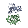
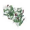
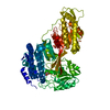
Title
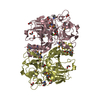
Crystal structure of predicted acetamidase/formamidase (YP_546212.1) from Methylobacillus flagellatus KT at 1.58 A resolution
Components
Twin-arginine translocation pathway signal protein
Keywords
HYDROLASE / YP_546212.1 / predicted acetamidase/formamidase / Acetamidase/Formamidase family / Structural Genomics / Joint Center for Structural Genomics / JCSG / Protein Structure Initiative / PSI-2
Function / homology
Acetamidase/Formamidase / Acetamidase/Formamidase family / hydrolase activity, acting on carbon-nitrogen (but not peptide) bonds, in linear amides / metal ion binding / Twin-arginine translocation pathway signal
Function and homology information
Biological species
Methylobacillus flagellatus KT (bacteria)
Method
X-RAY DIFFRACTION / SYNCHROTRON / SAD / Resolution: 1.58 Å
SEQUENCE 1. THE CONSTRUCT WAS EXPRESSED WITH A PURIFICATION TAG MGSDKIHHHHHHENLYFQG. THE TAG WAS ... SEQUENCE 1. THE CONSTRUCT WAS EXPRESSED WITH A PURIFICATION TAG MGSDKIHHHHHHENLYFQG. THE TAG WAS REMOVED WITH TEV PROTEASE LEAVING ONLY A GLYCINE (0) FOLLOWED BY THE TARGET SEQUENCE. 2. A PUTATIVE TWIN ARGININE SIGNAL PEPTIDE CLEAVAGE SITE IS LOCATED BETWEEN RESIDUES 57 AND 58. MASS SPECTROMETRY INDICATED THAT THE FULL LENGTH PROTEIN WAS PRESENT DURING PURIFICATION, HOWEVER, IT IS POSSIBLE THAT THE SHORTER CLEAVED PRODUCT WAS CRYSTALLIZED.
A: Twin-arginine translocation pathway signal protein B: Twin-arginine translocation pathway signal protein C: Twin-arginine translocation pathway signal protein D: Twin-arginine translocation pathway signal protein hetero molecules
Mass: 18.015 Da / Num. of mol.: 1696 / Source method: isolated from a natural source / Formula: H2O
Has protein modification
Y
Sequence details
REMARK 999 REMARK 999 SEQUENCE: THE CONSTRUCT WAS EXPRESSED WITH A PURIFICATION REMARK 999 TAG ...REMARK 999 REMARK 999 SEQUENCE: THE CONSTRUCT WAS EXPRESSED WITH A PURIFICATION REMARK 999 TAG MGSDKIHHHHHHENLYFQG. THE TAG WAS REMOVED WITH TEV PROTEASE REMARK 999 LEAVING ONLY A GLYCINE (0) FOLLOWED BY THE TARGET SEQUENCE.
-
Experimental details
-
Experiment
Experiment
Method: X-RAY DIFFRACTION / Number of used crystals: 1
-
Sample preparation
Crystal
Density Matthews: 1.8 Å3/Da / Density % sol: 31.83 %
Crystal grow
Temperature: 293 K / Method: vapor diffusion, sitting drop / pH: 6 Details: NANODROP, 1.0M LiCl, 20.0% PEG 6000, 0.1M MES pH 6.0, VAPOR DIFFUSION, SITTING DROP, temperature 293K
-
Data collection
Diffraction
Mean temperature: 100 K
Diffraction source
Source: SYNCHROTRON / Site: ALS / Beamline: 8.2.2 / Wavelength: 0.9796 Å
Monochromator: Double crystal Si(111) / Protocol: SINGLE WAVELENGTH / Monochromatic (M) / Laue (L): M / Scattering type: x-ray
Radiation wavelength
Wavelength: 0.9796 Å / Relative weight: 1
Reflection
Resolution: 1.58→29.814 Å / Num. obs: 196551 / % possible obs: 95.8 % / Redundancy: 1.9 % / Biso Wilson estimate: 13.67 Å2 / Rmerge(I) obs: 0.09 / Rsym value: 0.09 / Net I/σ(I): 7.2
Reflection shell
Diffraction-ID: 1
Resolution (Å)
Redundancy (%)
Rmerge(I) obs
Mean I/σ(I) obs
Num. measured all
Num. unique all
Rsym value
% possible all
1.58-1.62
1.9
0.459
1.6
27870
14334
0.459
94.2
1.62-1.67
1.9
0.384
2
27156
13960
0.384
94.4
1.67-1.71
1.9
0.323
2.3
26465
13607
0.323
94.6
1.71-1.77
1.9
0.272
2.8
25933
13334
0.272
95
1.77-1.82
1.9
0.217
3.5
25047
12910
0.217
95.2
1.82-1.89
1.9
0.177
4.1
24179
12471
0.177
95.3
1.89-1.96
1.9
0.146
4.9
23392
12076
0.146
95.6
1.96-2.04
1.9
0.122
6
22588
11682
0.122
95.8
2.04-2.13
1.9
0.103
6.9
21586
11198
0.103
96.1
2.13-2.23
1.9
0.093
7.4
20703
10744
0.093
96.2
2.23-2.36
1.9
0.089
7.7
19581
10220
0.089
96.4
2.36-2.5
1.9
0.087
7.8
18460
9674
0.087
96.5
2.5-2.67
1.9
0.083
7.9
17391
9108
0.083
96.6
2.67-2.88
1.9
0.078
8.3
16134
8465
0.078
96.3
2.88-3.16
1.9
0.067
9.5
14530
7774
0.067
96.5
3.16-3.53
1.8
0.058
11
12764
7039
0.058
96.6
3.53-4.08
1.8
0.052
12
10891
6196
0.052
96.1
4.08-5
2
0.052
12
10634
5342
0.052
98.7
5-7.07
2
0.07
9.2
8339
4182
0.07
99
7.07-29.814
2
0.074
8.4
4436
2235
0.074
97.8
-
Phasing
Phasing
Method: SAD
-
Processing
Software
Name
Version
Classification
NB
REFMAC
5.2.0019
refinement
PHENIX
refinement
SOLVE
phasing
MolProbity
3beta29
modelbuilding
SCALA
datascaling
PDB_EXTRACT
3
dataextraction
ADSC
Quantum
datacollection
MOSFLM
datareduction
Refinement
Method to determine structure: SAD / Resolution: 1.58→29.814 Å / Cor.coef. Fo:Fc: 0.975 / Cor.coef. Fo:Fc free: 0.959 / SU B: 3.099 / SU ML: 0.055 / TLS residual ADP flag: LIKELY RESIDUAL / Cross valid method: THROUGHOUT / σ(F): 0 / ESU R: 0.077 / ESU R Free: 0.082 Stereochemistry target values: MAXIMUM LIKELIHOOD WITH PHASES Details: 1. HYDROGENS HAVE BEEN ADDED IN THE RIDING POSITIONS. 2. ATOM RECORDS CONTAIN RESIDUAL B FACTORS ONLY. 3. A MET-INHIBITION PROTOCOL WAS USED FOR SELENOMETHIONINE INCORPORATION DURING PROTEIN ...Details: 1. HYDROGENS HAVE BEEN ADDED IN THE RIDING POSITIONS. 2. ATOM RECORDS CONTAIN RESIDUAL B FACTORS ONLY. 3. A MET-INHIBITION PROTOCOL WAS USED FOR SELENOMETHIONINE INCORPORATION DURING PROTEIN EXPRESSION. THE OCCUPANCY OF THE SE ATOMS IN THE MSE RESIDUES WAS REDUCED TO 0.75 FOR THE REDUCED SCATTERING POWER DUE TO PARTIAL S-MET INCORPORATION. 4. EDO, CL MOLECULES FROM THE CRYSTALLIZATION/CRYO SOLUTION ARE MODELED. 5. MG IONS ARE MODELED BASED ON DENSITY AND COORDINATION. THE ASSIGNMENT OF MG AND THE EDO NEXT TO IT IS TENTATIVE. 6. THERE ARE NO DENSITIES FOR THE N-TERMINAL RESIDUES 0-63 OF A/C/D CHAINS AND 0-67 OF B CHAIN ALTHOUGH MASS SPECTROSCOPY INDICATED THAT THEY ARE LIKELY TO BE PRESENT DURING PURIFICATION. THE SEQUENCE ANALYSIS INDICATED THAT THIS PROTEIN LIKELY CONTAINS A TWIN ARGININE SIGNAL PEPTIDE WITH A PUTATIVE CLEAVAGE SITE BETWEEN RESIDUES 57 AND 58. AS A RESULT, IT IS ALSO POSSIBLE THAT THE SHORTER CLEAVED PRODUCT WAS CRYSTALLIZED. 7. RAMACHANDRAN OUTLIERS 349 FOR ALL CHAINS ARE SUPPORTED BY WELL DEFINED DENSITY.
Rfactor
Num. reflection
% reflection
Selection details
Rfree
0.177
9907
5 %
RANDOM
Rwork
0.138
-
-
-
obs
0.14
196547
95.77 %
-
Solvent computation
Ion probe radii: 0.8 Å / Shrinkage radii: 0.8 Å / VDW probe radii: 1.2 Å / Solvent model: BABINET MODEL WITH MASK
Displacement parameters
Biso mean: 13.157 Å2
Baniso -1
Baniso -2
Baniso -3
1-
0.29 Å2
0.25 Å2
0.34 Å2
2-
-
0.37 Å2
-0.12 Å2
3-
-
-
-0.49 Å2
Refinement step
Cycle: LAST / Resolution: 1.58→29.814 Å
Protein
Nucleic acid
Ligand
Solvent
Total
Num. atoms
12906
0
174
1696
14776
Refine LS restraints
Refine-ID
Type
Dev ideal
Dev ideal target
Number
X-RAY DIFFRACTION
r_bond_refined_d
0.014
0.022
13635
X-RAY DIFFRACTION
r_bond_other_d
0.002
0.02
9295
X-RAY DIFFRACTION
r_angle_refined_deg
1.508
1.95
18518
X-RAY DIFFRACTION
r_angle_other_deg
0.929
3
22687
X-RAY DIFFRACTION
r_dihedral_angle_1_deg
6.464
5
1753
X-RAY DIFFRACTION
r_dihedral_angle_2_deg
30.972
24.133
600
X-RAY DIFFRACTION
r_dihedral_angle_3_deg
11.291
15
2144
X-RAY DIFFRACTION
r_dihedral_angle_4_deg
16.083
15
62
X-RAY DIFFRACTION
r_chiral_restr
0.095
0.2
1980
X-RAY DIFFRACTION
r_gen_planes_refined
0.007
0.02
15325
X-RAY DIFFRACTION
r_gen_planes_other
0.001
0.02
2759
X-RAY DIFFRACTION
r_nbd_refined
0.22
0.2
2537
X-RAY DIFFRACTION
r_nbd_other
0.204
0.2
10012
X-RAY DIFFRACTION
r_nbtor_refined
0.179
0.2
6590
X-RAY DIFFRACTION
r_nbtor_other
0.087
0.2
6881
X-RAY DIFFRACTION
r_xyhbond_nbd_refined
0.145
0.2
1291
X-RAY DIFFRACTION
r_metal_ion_refined
0.174
0.2
8
X-RAY DIFFRACTION
r_symmetry_vdw_refined
0.211
0.2
19
X-RAY DIFFRACTION
r_symmetry_vdw_other
0.223
0.2
61
X-RAY DIFFRACTION
r_symmetry_hbond_refined
0.145
0.2
38
X-RAY DIFFRACTION
r_mcbond_it
1.824
3
8940
X-RAY DIFFRACTION
r_mcbond_other
0.736
3
3448
X-RAY DIFFRACTION
r_mcangle_it
2.296
5
13607
X-RAY DIFFRACTION
r_scbond_it
4.01
8
5808
X-RAY DIFFRACTION
r_scangle_it
5.052
11
4866
Refine LS restraints NCS
Ens-ID: 1 / Refine-ID: X-RAY DIFFRACTION
Dom-ID
Auth asym-ID
Number
Type
Rms dev position (Å)
Weight position
1
A
2357
MEDIUMPOSITIONAL
0.12
0.5
2
B
2357
MEDIUMPOSITIONAL
0.18
0.5
3
C
2357
MEDIUMPOSITIONAL
0.1
0.5
4
D
2357
MEDIUMPOSITIONAL
0.09
0.5
1
A
2748
LOOSEPOSITIONAL
0.18
5
2
B
2748
LOOSEPOSITIONAL
0.25
5
3
C
2748
LOOSEPOSITIONAL
0.3
5
4
D
2748
LOOSEPOSITIONAL
0.18
5
1
A
2357
MEDIUMTHERMAL
1.25
2
2
B
2357
MEDIUMTHERMAL
0.92
2
3
C
2357
MEDIUMTHERMAL
1.3
2
4
D
2357
MEDIUMTHERMAL
0.88
2
1
A
2748
LOOSETHERMAL
1.85
10
2
B
2748
LOOSETHERMAL
1.55
10
3
C
2748
LOOSETHERMAL
1.94
10
4
D
2748
LOOSETHERMAL
1.42
10
LS refinement shell
Resolution: 1.58→1.621 Å / Total num. of bins used: 20
Rfactor
Num. reflection
% reflection
Rfree
0.276
705
-
Rwork
0.214
13595
-
all
-
14300
-
obs
-
-
94.25 %
Refinement TLS params.
Method: refined / Refine-ID: X-RAY DIFFRACTION
ID
L11 (°2)
L12 (°2)
L13 (°2)
L22 (°2)
L23 (°2)
L33 (°2)
S11 (Å °)
S12 (Å °)
S13 (Å °)
S21 (Å °)
S22 (Å °)
S23 (Å °)
S31 (Å °)
S32 (Å °)
S33 (Å °)
T11 (Å2)
T12 (Å2)
T13 (Å2)
T22 (Å2)
T23 (Å2)
T33 (Å2)
Origin x (Å)
Origin y (Å)
Origin z (Å)
1
0.3478
-0.0627
0.0089
0.2158
0.095
0.4993
-0.0612
-0.0365
-0.0837
0.0653
-0.0172
0.0132
0.164
0.0431
0.0784
0.0337
0.0182
0.0363
-0.0517
0.0264
-0.003
19.0798
37.1237
11.9073
2
0.3407
-0.0672
0.0285
0.3018
-0.0501
0.2955
-0.0228
0.0771
-0.0219
-0.0699
-0.0339
0.007
0.0606
-0.0114
0.0568
-0.0041
-0.0066
0.0082
-0.0068
-0.0362
-0.0302
8.0207
47.1373
-22.2396
3
0.3452
-0.0132
-0.0975
0.1987
-0.0535
0.2219
-0.0075
-0.0399
0.0011
0.0352
-0.0112
0.0017
-0.0075
0.015
0.0187
-0.0315
-0.0031
-0.0173
-0.0276
-0.0109
-0.0287
7.2239
71.971
21.7988
4
0.3551
-0.0142
-0.0918
0.141
0.0348
0.2826
0.0145
0.068
0.0595
-0.0237
-0.0208
-0.0107
-0.0332
-0.0033
0.0063
-0.0328
0.0068
-0.0145
-0.0227
0.025
-0.0123
20.968
82.0128
-11.5192
Refinement TLS group
ID
Refine-ID
Refine TLS-ID
Auth asym-ID
Label asym-ID
Auth seq-ID
Label seq-ID
1
X-RAY DIFFRACTION
1
A
A
64 - 482
65 - 483
2
X-RAY DIFFRACTION
2
B
B
68 - 481
69 - 482
3
X-RAY DIFFRACTION
3
C
C
64 - 482
65 - 483
4
X-RAY DIFFRACTION
4
D
D
68 - 481
69 - 482
+
About Yorodumi
-
News
-
Feb 9, 2022. New format data for meta-information of EMDB entries
New format data for meta-information of EMDB entries
Version 3 of the EMDB header file is now the official format.
The previous official version 1.9 will be removed from the archive.
In the structure databanks used in Yorodumi, some data are registered as the other names, "COVID-19 virus" and "2019-nCoV". Here are the details of the virus and the list of structure data.
Jan 31, 2019. EMDB accession codes are about to change! (news from PDBe EMDB page)
EMDB accession codes are about to change! (news from PDBe EMDB page)
The allocation of 4 digits for EMDB accession codes will soon come to an end. Whilst these codes will remain in use, new EMDB accession codes will include an additional digit and will expand incrementally as the available range of codes is exhausted. The current 4-digit format prefixed with “EMD-” (i.e. EMD-XXXX) will advance to a 5-digit format (i.e. EMD-XXXXX), and so on. It is currently estimated that the 4-digit codes will be depleted around Spring 2019, at which point the 5-digit format will come into force.
The EM Navigator/Yorodumi systems omit the EMD- prefix.
Related info.:Q: What is EMD? / ID/Accession-code notation in Yorodumi/EM Navigator
Yorodumi is a browser for structure data from EMDB, PDB, SASBDB, etc.
This page is also the successor to EM Navigator detail page, and also detail information page/front-end page for Omokage search.
The word "yorodu" (or yorozu) is an old Japanese word meaning "ten thousand". "mi" (miru) is to see.
Related info.:EMDB / PDB / SASBDB / Comparison of 3 databanks / Yorodumi Search / Aug 31, 2016. New EM Navigator & Yorodumi / Yorodumi Papers / Jmol/JSmol / Function and homology information / Changes in new EM Navigator and Yorodumi
 Movie
Movie Controller

 Yorodumi
 Open data
 Basic information
 Components
 Keywords
 Function and homology information
 Methylobacillus flagellatus KT (bacteria)
 X-RAY DIFFRACTION /
  Authors
 Citation
 Structure visualization
 Downloads & links
 Other downloads

 PDBj
PDBj Assembly


 Mass: 24.305 Da / Num. of mol.: 8 / Source method: obtained synthetically / Formula: Mg
 Mass: 35.453 Da / Num. of mol.: 2 / Source method: obtained synthetically / Formula: Cl
 Mass: 62.068 Da / Num. of mol.: 41 / Source method: obtained synthetically / Formula: C2H6O2
 Mass: 18.015 Da / Num. of mol.: 1696 / Source method: isolated from a natural source / Formula: H2O
 Sample preparation
 / Beamline: 8.2.2 / Wavelength: 0.9796 Å
 Processing