 Movie
Movie Controller

[English] 日本語
 Yorodumi
- PDB-6f2v: Crystal structure of ectonucleotide phosphodiesterase/pyrophospha... -
+ Open data

- Basic information
| Entry | Database: PDB / ID: 6f2v | |||||||||
|---|---|---|---|---|---|---|---|---|---|---|
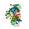
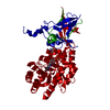
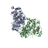
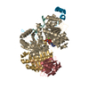
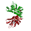
| Title | Crystal structure of ectonucleotide phosphodiesterase/pyrophosphatase-3 (NPP3) in complex with AMP | |||||||||
 Components Components | Ectonucleotide pyrophosphatase/phosphodiesterase family member 3 | |||||||||
 Keywords Keywords | HYDROLASE / enzyme / ectonucleotide phosphodiesterase/pyrophosphatase / complex / CYTOSOLIC PROTEIN | |||||||||
| Function / homology |  Function and homology information Function and homology informationGTP diphosphatase activity / cyclic-GMP-AMP hydrolase activity / bis(5'-adenosyl)-triphosphatase activity / negative regulation of mast cell activation involved in immune response / basophil activation involved in immune response / UTP diphosphatase activity / dinucleotide phosphatase activity / phosphodiesterase I / nucleotide diphosphatase / bis(5'-nucleosyl)-tetraphosphatase (asymmetrical) activity ...GTP diphosphatase activity / cyclic-GMP-AMP hydrolase activity / bis(5'-adenosyl)-triphosphatase activity / negative regulation of mast cell activation involved in immune response / basophil activation involved in immune response / UTP diphosphatase activity / dinucleotide phosphatase activity / phosphodiesterase I / nucleotide diphosphatase / bis(5'-nucleosyl)-tetraphosphatase (asymmetrical) activity / negative regulation of mast cell proliferation / bis(5'-adenosyl)-pentaphosphatase activity / nucleoside triphosphate catabolic process / regulation of smooth muscle cell differentiation / pyrimidine nucleotide metabolic process / nucleoside triphosphate diphosphatase activity / ATP diphosphatase activity / phosphate ion homeostasis / phosphate-containing compound metabolic process / phosphodiesterase I activity / negative regulation of cGAS/STING signaling pathway / ATP metabolic process / Hydrolases; Acting on acid anhydrides; In phosphorus-containing anhydrides / negative regulation of inflammatory response / nucleic acid binding / apical plasma membrane / external side of plasma membrane / calcium ion binding / perinuclear region of cytoplasm / cell surface / extracellular region / zinc ion binding / ATP binding / plasma membrane Similarity search - Function | |||||||||
| Biological species |  | |||||||||
| Method |  X-RAY DIFFRACTION / SYNCHROTRON / MOLECULAR REPLACEMENT / Resolution: 2.5 Å X-RAY DIFFRACTION / SYNCHROTRON / MOLECULAR REPLACEMENT / Resolution: 2.5 Å | |||||||||
 Authors Authors | Dohler, C. / Zebisch, M. / Strater, N. | |||||||||
| Funding support |  Germany, 1items Germany, 1items
| |||||||||
 Citation Citation | Journal: Sci Rep / Year: 2018 Title: Crystal structure and substrate binding mode of ectonucleotide phosphodiesterase/pyrophosphatase-3 (NPP3). Authors: Dohler, C. / Zebisch, M. / Strater, N. | |||||||||
| History |
|
- Structure visualization
| Structure viewer | Molecule: MolmilJmol/JSmol |
|---|
- Downloads & links
-Download
| PDBx/mmCIF format | 6f2v.cif.gz | 611.3 KB | Display | PDBx/mmCIF format |
|---|---|---|---|---|
| PDB format | pdb6f2v.ent.gz | 500.9 KB | Display | PDB format |
| PDBx/mmJSON format | 6f2v.json.gz | Tree view | PDBx/mmJSON format | |
| Others |  Other downloads Other downloads |
-Validation report
| Arichive directory | https://data.pdbj.org/pub/pdb/validation_reports/f2/6f2vftp://data.pdbj.org/pub/pdb/validation_reports/f2/6f2v | HTTPS FTP |
|---|
-Related structure data
| Related structure data |  6f2tC  6f2yC  6f30C  6f33C  2xr9S S: Starting model for refinement C: citing same article ( |
|---|---|
| Similar structure data |


-Links
 PDBj
PDBj


- Assembly
| Deposited unit | 
| ||||||||
|---|---|---|---|---|---|---|---|---|---|
| 1 | 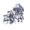
| ||||||||
| 2 | 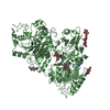
| ||||||||
| Unit cell |
|
-Components
-Protein , 1 types, 2 molecules AB
| #1: Protein | Mass: 85399.375 Da / Num. of mol.: 2 Source method: isolated from a genetically manipulated source Source: (gene. exp.)  Homo sapiens (human) Homo sapiens (human)References: UniProt: P97675, phosphodiesterase I, nucleotide diphosphatase |
|---|
-Sugars , 3 types, 10 molecules 
| #2: Polysaccharide | 2-acetamido-2-deoxy-beta-D-glucopyranose-(1-4)-2-acetamido-2-deoxy-beta-D-glucopyranose Source method: isolated from a genetically manipulated source #3: Polysaccharide | beta-D-mannopyranose-(1-4)-2-acetamido-2-deoxy-beta-D-glucopyranose-(1-4)-2-acetamido-2-deoxy-beta- ...beta-D-mannopyranose-(1-4)-2-acetamido-2-deoxy-beta-D-glucopyranose-(1-4)-2-acetamido-2-deoxy-beta-D-glucopyranose | Source method: isolated from a genetically manipulated source #7: Sugar | ChemComp-NAG /  Type: D-saccharide, beta linking / Mass: 221.208 Da / Num. of mol.: 5 Type: D-saccharide, beta linking / Mass: 221.208 Da / Num. of mol.: 5Source method: isolated from a genetically manipulated source Formula: C8H15NO6 |
|---|
-Non-polymers , 4 types, 472 molecules 


| #4: Chemical |  Mass: 347.221 Da / Num. of mol.: 2 / Source method: obtained synthetically / Formula: C10H14N5O7P / Comment: AMP*YM Mass: 347.221 Da / Num. of mol.: 2 / Source method: obtained synthetically / Formula: C10H14N5O7P / Comment: AMP*YM#5: Chemical | ChemComp-ZN /  Mass: 65.409 Da / Num. of mol.: 4 / Source method: obtained synthetically / Formula: Zn Mass: 65.409 Da / Num. of mol.: 4 / Source method: obtained synthetically / Formula: Zn#6: Chemical |  Mass: 40.078 Da / Num. of mol.: 2 / Source method: obtained synthetically / Formula: Ca Mass: 40.078 Da / Num. of mol.: 2 / Source method: obtained synthetically / Formula: Ca#8: Water | ChemComp-HOH / | Mass: 18.015 Da / Num. of mol.: 464 / Source method: isolated from a natural source / Formula: H2O |
|---|
-Details
| Has protein modification | Y |
|---|
-Experimental details
-Experiment
| Experiment | Method: X-RAY DIFFRACTION / Number of used crystals: 1 |
|---|
- Sample preparation
| Crystal | Density Matthews: 3.06 Å3/Da / Density % sol: 59.84 % |
|---|---|
| Crystal grow | Temperature: 292 K / Method: vapor diffusion, hanging drop / pH: 6 Details: 0.2 M KCl, 0.1 M MgAcetate, 0.05 NaCacodylate pH 6.0, 10.2% PEG8000, 1 mM CaCl2, 0.1 mM ZnSO4 |
-Data collection
| Diffraction | Mean temperature: 100 K |
|---|---|
| Diffraction source | Source: SYNCHROTRON / Site: BESSY / Beamline: 14.1 / Wavelength: 0.91841 Å |
| Detector | Type: DECTRIS PILATUS 6M / Detector: PIXEL / Date: Feb 16, 2013 |
| Radiation | Monochromator: SI(111) / Protocol: SINGLE WAVELENGTH / Monochromatic (M) / Laue (L): M / Scattering type: x-ray |
| Radiation wavelength | Wavelength: 0.91841 Å / Relative weight: 1 |
| Reflection | Resolution: 2.5→37.81 Å / Num. obs: 70943 / % possible obs: 99.8 % / Redundancy: 3.6 % / Biso Wilson estimate: 56.69 Å2 / Rpim(I) all: 0.097 / Net I/σ(I): 12.3 |
| Reflection shell | Resolution: 2.5→2.54 Å |
- Processing
| Software |
| |||||||||||||||||||||||||||||||||||||||||||||||||||||||||||||||||||||||||||||||||||||||||||||||||||||||||||||||||||||||||||||
|---|---|---|---|---|---|---|---|---|---|---|---|---|---|---|---|---|---|---|---|---|---|---|---|---|---|---|---|---|---|---|---|---|---|---|---|---|---|---|---|---|---|---|---|---|---|---|---|---|---|---|---|---|---|---|---|---|---|---|---|---|---|---|---|---|---|---|---|---|---|---|---|---|---|---|---|---|---|---|---|---|---|---|---|---|---|---|---|---|---|---|---|---|---|---|---|---|---|---|---|---|---|---|---|---|---|---|---|---|---|---|---|---|---|---|---|---|---|---|---|---|---|---|---|---|---|---|
| Refinement | Method to determine structure: MOLECULAR REPLACEMENT Starting model: 2XR9 Resolution: 2.5→37.81 Å / Cor.coef. Fo:Fc: 0.922 / Cor.coef. Fo:Fc free: 0.917 / Rfactor Rfree error: 0 / SU R Cruickshank DPI: 0.366 / Cross valid method: THROUGHOUT / σ(F): 0 / SU R Blow DPI: 0.355 / SU Rfree Blow DPI: 0.213 / SU Rfree Cruickshank DPI: 0.218
| |||||||||||||||||||||||||||||||||||||||||||||||||||||||||||||||||||||||||||||||||||||||||||||||||||||||||||||||||||||||||||||
| Displacement parameters | Biso mean: 54.37 Å2
| |||||||||||||||||||||||||||||||||||||||||||||||||||||||||||||||||||||||||||||||||||||||||||||||||||||||||||||||||||||||||||||
| Refine analyze | Luzzati coordinate error obs: 0.34 Å | |||||||||||||||||||||||||||||||||||||||||||||||||||||||||||||||||||||||||||||||||||||||||||||||||||||||||||||||||||||||||||||
| Refinement step | Cycle: 1 / Resolution: 2.5→37.81 Å
| |||||||||||||||||||||||||||||||||||||||||||||||||||||||||||||||||||||||||||||||||||||||||||||||||||||||||||||||||||||||||||||
| Refine LS restraints |
| |||||||||||||||||||||||||||||||||||||||||||||||||||||||||||||||||||||||||||||||||||||||||||||||||||||||||||||||||||||||||||||
| LS refinement shell | Resolution: 2.5→2.56 Å / Rfactor Rfree error: 0 / Total num. of bins used: 20
| |||||||||||||||||||||||||||||||||||||||||||||||||||||||||||||||||||||||||||||||||||||||||||||||||||||||||||||||||||||||||||||
| Refinement TLS params. | Method: refined / Refine-ID: X-RAY DIFFRACTION
| |||||||||||||||||||||||||||||||||||||||||||||||||||||||||||||||||||||||||||||||||||||||||||||||||||||||||||||||||||||||||||||
| Refinement TLS group |
|
