 Movie
Movie Controller

[English] 日本語
 Yorodumi
- PDB-3b1j: Crystal structure of Glyceraldehyde-3-Phosphate Dehydrogenase com... -
+ Open data

- Basic information
| Entry | Database: PDB / ID: 3b1j | ||||||
|---|---|---|---|---|---|---|---|
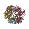
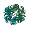
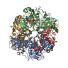
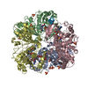
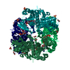
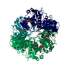
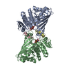
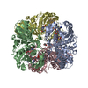
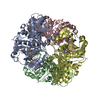
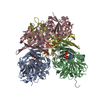
| Title | Crystal structure of Glyceraldehyde-3-Phosphate Dehydrogenase complexed with CP12 in the presence of copper from Synechococcus elongatus | ||||||
 Components Components |
| ||||||
 Keywords Keywords | OXIDOREDUCTASE/PROTEIN BINDING / alpha/beta fold / OXIDOREDUCTASE-PROTEIN BINDING complex | ||||||
| Function / homology |  Function and homology information Function and homology informationglyceraldehyde-3-phosphate dehydrogenase [NAD(P)+] (phosphorylating) activity / negative regulation of reductive pentose-phosphate cycle / Oxidoreductases; Acting on the aldehyde or oxo group of donors; With NAD+ or NADP+ as acceptor / glucose metabolic process / NAD binding / NADP binding / nucleotide binding / metal ion binding Similarity search - Function | ||||||
| Biological species |  Synechococcus elongatus (bacteria) Synechococcus elongatus (bacteria) | ||||||
| Method |  X-RAY DIFFRACTION / SYNCHROTRON / MOLECULAR REPLACEMENT / Resolution: 2.2 Å X-RAY DIFFRACTION / SYNCHROTRON / MOLECULAR REPLACEMENT / Resolution: 2.2 Å | ||||||
 Authors Authors | Matsumura, H. / Kai, A. / Inoue, T. | ||||||
 Citation Citation | Journal: Structure / Year: 2011 Title: Structure Basis for the Regulation of Glyceraldehyde-3-Phosphate Dehydrogenase Activity via the Intrinsically Disordered Protein CP12. Authors: Matsumura, H. / Kai, A. / Maeda, T. / Tamoi, M. / Satoh, A. / Tamura, H. / Hirose, M. / Ogawa, T. / Kizu, N. / Wadano, A. / Inoue, T. / Shigeoka, S. | ||||||
| History |
|
- Structure visualization
| Structure viewer | Molecule: MolmilJmol/JSmol |
|---|
- Downloads & links
-Download
| PDBx/mmCIF format | 3b1j.cif.gz | 155.1 KB | Display | PDBx/mmCIF format |
|---|---|---|---|---|
| PDB format | pdb3b1j.ent.gz | 122.9 KB | Display | PDB format |
| PDBx/mmJSON format | 3b1j.json.gz | Tree view | PDBx/mmJSON format | |
| Others |  Other downloads Other downloads |
-Validation report
| Arichive directory | https://data.pdbj.org/pub/pdb/validation_reports/b1/3b1jftp://data.pdbj.org/pub/pdb/validation_reports/b1/3b1j | HTTPS FTP |
|---|
-Related structure data
| Related structure data |  3b1kC  3b20C 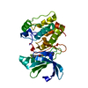 3d2iS C: citing same article ( S: Starting model for refinement |
|---|---|
| Similar structure data |
-Links
 PDBj
PDBj
- Assembly
| Deposited unit | 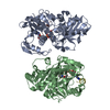
| ||||||||
|---|---|---|---|---|---|---|---|---|---|
| 1 | 
| ||||||||
| Unit cell |
|
-Components
| #1: Protein | Mass: 37032.070 Da / Num. of mol.: 2 Source method: isolated from a genetically manipulated source Source: (gene. exp.) Synechococcus elongatus (bacteria) / Strain: PCC 7942 / Gene: gap2, Synpcc7942_1742 / Production host: References: UniProt: Q9R6W2, glyceraldehyde-3-phosphate dehydrogenase (NADP+) (phosphorylating) #2: Protein/peptide | Mass: 2830.917 Da / Num. of mol.: 2 / Fragment: UNP RESIDUES 51-75 Source method: isolated from a genetically manipulated source Source: (gene. exp.) Synechococcus elongatus (bacteria) / Strain: PCC 7942 / Gene: cp12, Synpcc7942_0361 / Production host: #3: Chemical |   Mass: 663.425 Da / Num. of mol.: 2 / Source method: obtained synthetically / Formula: C21H27N7O14P2 / Comment: NAD*YM Mass: 663.425 Da / Num. of mol.: 2 / Source method: obtained synthetically / Formula: C21H27N7O14P2 / Comment: NAD*YM#4: Chemical |   Mass: 63.546 Da / Num. of mol.: 2 / Source method: obtained synthetically / Formula: Cu Mass: 63.546 Da / Num. of mol.: 2 / Source method: obtained synthetically / Formula: Cu#5: Water | ChemComp-HOH / |  Mass: 18.015 Da / Num. of mol.: 210 / Source method: isolated from a natural source / Formula: H2O Mass: 18.015 Da / Num. of mol.: 210 / Source method: isolated from a natural source / Formula: H2OHas protein modification | Y | |
|---|
-Experimental details
-Experiment
| Experiment | Method: X-RAY DIFFRACTION / Number of used crystals: 1 |
|---|
- Sample preparation
| Crystal | Density Matthews: 2.6 Å3/Da / Density % sol: 52.78 % |
|---|---|
| Crystal grow | Temperature: 293 K / Method: vapor diffusion, hanging drop / pH: 7.5 Details: 20%(v/v) PEG3350, 0.2M magnesium acetate, pH 7.5, VAPOR DIFFUSION, HANGING DROP, temperature 293K |
-Data collection
| Diffraction | Mean temperature: 100 K |
|---|---|
| Diffraction source | Source: SYNCHROTRON / Site: SPring-8  / Beamline: BL41XU / Wavelength: 1 Å / Beamline: BL41XU / Wavelength: 1 Å |
| Detector | Date: Dec 7, 2009 |
| Radiation | Monochromator: Si 111 CHANNEL / Protocol: SINGLE WAVELENGTH / Monochromatic (M) / Laue (L): M / Scattering type: x-ray |
| Radiation wavelength | Wavelength: 1 Å / Relative weight: 1 |
| Reflection | Resolution: 2.2→50 Å / Num. all: 42240 / Num. obs: 42240 / % possible obs: 99.8 % / Observed criterion σ(I): 0.187 / Rmerge(I) obs: 0.105 |
| Reflection shell | Resolution: 2.2→2.28 Å / Rmerge(I) obs: 0.435 / % possible all: 99.7 |
- Processing
| Software |
| ||||||||||||||||||||
|---|---|---|---|---|---|---|---|---|---|---|---|---|---|---|---|---|---|---|---|---|---|
| Refinement | Method to determine structure: MOLECULAR REPLACEMENT Starting model: 3D2I Resolution: 2.2→50 Å / σ(F): 0 / Stereochemistry target values: Engh & Huber
| ||||||||||||||||||||
| Refinement step | Cycle: LAST / Resolution: 2.2→50 Å
| ||||||||||||||||||||
| Refine LS restraints |
|
