 Movie
Movie Controller

[English] 日本語
 Yorodumi
- PDB-3b09: Crystal structure of the N-domain of FKBP22 from Shewanella sp. SIB1 -
+ Open data

- Basic information
| Entry | Database: PDB / ID: 3b09 | ||||||
|---|---|---|---|---|---|---|---|
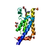
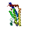
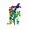
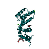
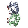
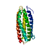
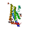
| Title | Crystal structure of the N-domain of FKBP22 from Shewanella sp. SIB1 | ||||||
 Components Components | Peptidyl-prolyl cis-trans isomerase | ||||||
 Keywords Keywords | CHAPERONE / Val-Leu zipper / helices | ||||||
| Function / homology |  Function and homology information Function and homology informationpeptidylprolyl isomerase / peptidyl-prolyl cis-trans isomerase activity / protein folding Similarity search - Function | ||||||
| Biological species |  Shewanella (bacteria) Shewanella (bacteria) | ||||||
| Method |  X-RAY DIFFRACTION / SYNCHROTRON / MAD / Resolution: 1.9 Å X-RAY DIFFRACTION / SYNCHROTRON / MAD / Resolution: 1.9 Å | ||||||
 Authors Authors | Budiman, C. / Angkawidjaja, C. / Motoike, H. / Koga, Y. / Takano, K. / Kanaya, S. | ||||||
 Citation Citation | Journal: Protein Sci. / Year: 2011 Title: Crystal structure of N-domain of FKBP22 from Shewanella sp. SIB1: dimer dissociation by disruption of Val-Leu knot Authors: Budiman, C. / Angkawidjaja, C. / Motoike, H. / Koga, Y. / Takano, K. / Kanaya, S. | ||||||
| History |
|
- Structure visualization
| Structure viewer | Molecule: MolmilJmol/JSmol |
|---|
- Downloads & links
-Download
| PDBx/mmCIF format | 3b09.cif.gz | 25.6 KB | Display | PDBx/mmCIF format |
|---|---|---|---|---|
| PDB format | pdb3b09.ent.gz | 16.1 KB | Display | PDB format |
| PDBx/mmJSON format | 3b09.json.gz | Tree view | PDBx/mmJSON format | |
| Others |  Other downloads Other downloads |
-Validation report
| Arichive directory | https://data.pdbj.org/pub/pdb/validation_reports/b0/3b09ftp://data.pdbj.org/pub/pdb/validation_reports/b0/3b09 | HTTPS FTP |
|---|
-Related structure data
| Similar structure data |
|---|


-Links
 PDBj
PDBj


- Assembly
| Deposited unit | 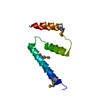
| ||||||||
|---|---|---|---|---|---|---|---|---|---|
| 1 | 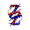
| ||||||||
| Unit cell |
|
-Components
| #1: Protein | Mass: 9706.961 Da / Num. of mol.: 1 / Fragment: N-terminal domain, residues 1-68 Source method: isolated from a genetically manipulated source Source: (gene. exp.) Shewanella (bacteria) / Strain: SIB1 / Gene: fklB / Plasmid: pET28a / Production host: |
|---|---|
| #2: Water | ChemComp-HOH /  Mass: 18.015 Da / Num. of mol.: 28 / Source method: isolated from a natural source / Formula: H2O Mass: 18.015 Da / Num. of mol.: 28 / Source method: isolated from a natural source / Formula: H2O |
| Has protein modification | Y |
-Experimental details
-Experiment
| Experiment | Method: X-RAY DIFFRACTION / Number of used crystals: 1 |
|---|
- Sample preparation
| Crystal | Density Matthews: 2.38 Å3/Da / Density % sol: 48.35 % |
|---|---|
| Crystal grow | Temperature: 277 K / Method: vapor diffusion, sitting drop / pH: 6.2 Details: 0.1M Na/K Phosphate, 20% PEG 1000, 0.2 M NaCl, pH 6.2, VAPOR DIFFUSION, SITTING DROP, temperature 277K |
-Data collection
| Diffraction | Mean temperature: 100 K | ||||||||||||
|---|---|---|---|---|---|---|---|---|---|---|---|---|---|
| Diffraction source | Source: SYNCHROTRON / Site: SPring-8  / Beamline: BL44B2 / Wavelength: 0.97888, 0.97919, 0.96500 / Beamline: BL44B2 / Wavelength: 0.97888, 0.97919, 0.96500 | ||||||||||||
| Detector | Type: MARMOSAIC 225 mm CCD / Detector: CCD / Date: Jul 15, 2010 / Details: mirrors | ||||||||||||
| Radiation | Monochromator: mirrors / Protocol: MAD / Monochromatic (M) / Laue (L): M / Scattering type: x-ray | ||||||||||||
| Radiation wavelength |
| ||||||||||||
| Reflection | Resolution: 1.9→50 Å / Num. obs: 7712 / % possible obs: 98.3 % / Observed criterion σ(F): 5 / Observed criterion σ(I): 5 / Redundancy: 5.4 % / Rmerge(I) obs: 0.114 / Rsym value: 0.119 / Net I/σ(I): 18.2 | ||||||||||||
| Reflection shell | Resolution: 1.9→1.93 Å / Redundancy: 5.3 % / Rmerge(I) obs: 0.423 / Mean I/σ(I) obs: 2.6 / Num. unique all: 364 / Rsym value: 0.398 / % possible all: 100 |
- Processing
| Software |
| |||||||||||||||||||||||||||||||||||||||||||||||||||||||||||||||||
|---|---|---|---|---|---|---|---|---|---|---|---|---|---|---|---|---|---|---|---|---|---|---|---|---|---|---|---|---|---|---|---|---|---|---|---|---|---|---|---|---|---|---|---|---|---|---|---|---|---|---|---|---|---|---|---|---|---|---|---|---|---|---|---|---|---|---|
| Refinement | Method to determine structure: MAD / Resolution: 1.9→34.38 Å / Cor.coef. Fo:Fc: 0.927 / Cor.coef. Fo:Fc free: 0.89 / SU B: 2.996 / SU ML: 0.091 / Cross valid method: THROUGHOUT / ESU R Free: 0.143 / Stereochemistry target values: MAXIMUM LIKELIHOOD / Details: HYDROGENS HAVE BEEN ADDED IN THE RIDING POSITIONS
| |||||||||||||||||||||||||||||||||||||||||||||||||||||||||||||||||
| Solvent computation | Ion probe radii: 0.8 Å / Shrinkage radii: 0.8 Å / VDW probe radii: 1.4 Å / Solvent model: MASK | |||||||||||||||||||||||||||||||||||||||||||||||||||||||||||||||||
| Displacement parameters | Biso mean: 30.92 Å2
| |||||||||||||||||||||||||||||||||||||||||||||||||||||||||||||||||
| Refinement step | Cycle: LAST / Resolution: 1.9→34.38 Å
| |||||||||||||||||||||||||||||||||||||||||||||||||||||||||||||||||
| Refine LS restraints |
| |||||||||||||||||||||||||||||||||||||||||||||||||||||||||||||||||
| LS refinement shell | Resolution: 1.899→1.948 Å / Total num. of bins used: 20
|
