 Movie
Movie Controller

[English] 日本語
 Yorodumi
- PDB-3axe: The truncated Fibrobacter succinogenes 1,3-1,4-beta-D-glucanase V... -
+ Open data

- Basic information
| Entry | Database: PDB / ID: 3axe | |||||||||
|---|---|---|---|---|---|---|---|---|---|---|
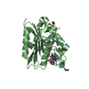
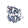
| Title | The truncated Fibrobacter succinogenes 1,3-1,4-beta-D-glucanase V18Y/W203Y in complex with cellotetraose (cellobiose density was observed) | |||||||||
 Components Components | Beta-glucanase | |||||||||
 Keywords Keywords | HYDROLASE / glucanase / cellobiose/cellotetraose | |||||||||
| Function / homology |  Function and homology information Function and homology information | |||||||||
| Biological species |  Fibrobacter succinogenes (bacteria) Fibrobacter succinogenes (bacteria) | |||||||||
| Method |  X-RAY DIFFRACTION / SYNCHROTRON / MOLECULAR REPLACEMENT / Resolution: 1.53 Å X-RAY DIFFRACTION / SYNCHROTRON / MOLECULAR REPLACEMENT / Resolution: 1.53 Å | |||||||||
 Authors Authors | Huang, J.W. / Cheng, Y.S. / Ko, T.P. / Lin, C.Y. / Lai, H.L. / Chen, C.C. / Ma, Y. / Huang, C.H. / Zheng, Y. / Liu, J.R. / Guo, R.T. | |||||||||
 Citation Citation | Journal: Appl.Microbiol.Biotechnol. / Year: 2012 Title: Rational design to improve thermostability and specific activity of the truncated Fibrobacter succinogenes 1,3-1,4-beta-D-glucanase Authors: Huang, J.W. / Cheng, Y.S. / Ko, T.P. / Lin, C.Y. / Lai, H.L. / Chen, C.C. / Ma, Y. / Zheng, Y. / Huang, C.H. / Zou, P. / Liu, J.R. / Guo, R.T. | |||||||||
| History |
|
- Structure visualization
| Structure viewer | Molecule: MolmilJmol/JSmol |
|---|
- Downloads & links
-Download
| PDBx/mmCIF format | 3axe.cif.gz | 71.9 KB | Display | PDBx/mmCIF format |
|---|---|---|---|---|
| PDB format | pdb3axe.ent.gz | 49.9 KB | Display | PDB format |
| PDBx/mmJSON format | 3axe.json.gz | Tree view | PDBx/mmJSON format | |
| Others |  Other downloads Other downloads |
-Validation report
| Arichive directory | https://data.pdbj.org/pub/pdb/validation_reports/ax/3axeftp://data.pdbj.org/pub/pdb/validation_reports/ax/3axe | HTTPS FTP |
|---|
-Related structure data
| Related structure data |  3axdC  1zm1S C: citing same article ( S: Starting model for refinement |
|---|---|
| Similar structure data |


-Links
 PDBj
PDBj
- Assembly
| Deposited unit | 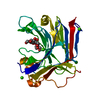
| ||||||||
|---|---|---|---|---|---|---|---|---|---|
| 1 |
| ||||||||
| Unit cell |
|
-Components
| #1: Protein | Mass: 27827.656 Da / Num. of mol.: 1 / Fragment: UNP RESIDUES 25-271 / Mutation: V18Y, W203Y Source method: isolated from a genetically manipulated source Source: (gene. exp.) Fibrobacter succinogenes (bacteria) / Strain: ATCC 19169, S85 / Gene: Fisuc_2961, FSU_0226 / Plasmid: pET 32 Xa/LIC / Production host: | ||
|---|---|---|---|
| #2: Polysaccharide | beta-D-glucopyranose-(1-4)-beta-D-glucopyranose / beta-cellobiose  Source method: isolated from a genetically manipulated source Details: oligosaccharide / References: beta-cellobiose | ||
| #3: Chemical | ChemComp-TRS /   Mass: 122.143 Da / Num. of mol.: 1 / Source method: obtained synthetically / Formula: C4H12NO3 / Comment: pH buffer*YM Mass: 122.143 Da / Num. of mol.: 1 / Source method: obtained synthetically / Formula: C4H12NO3 / Comment: pH buffer*YM | ||
| #4: Chemical | ChemComp-CA /   Mass: 40.078 Da / Num. of mol.: 4 / Source method: obtained synthetically / Formula: Ca Mass: 40.078 Da / Num. of mol.: 4 / Source method: obtained synthetically / Formula: Ca#5: Water | ChemComp-HOH / |  Mass: 18.015 Da / Num. of mol.: 332 / Source method: isolated from a natural source / Formula: H2O Mass: 18.015 Da / Num. of mol.: 332 / Source method: isolated from a natural source / Formula: H2O |
-Experimental details
-Experiment
| Experiment | Method: X-RAY DIFFRACTION / Number of used crystals: 1 |
|---|
- Sample preparation
| Crystal | Density Matthews: 1.94 Å3/Da / Density % sol: 36.67 % |
|---|---|
| Crystal grow | Temperature: 298 K / Method: vapor diffusion, sitting drop / pH: 7.5 Details: 0.15M Tris, pH 8.5, 0.4M Calcium acetate, 33% PEG 5KMME, VAPOR DIFFUSION, SITTING DROP, temperature 298.0K |
-Data collection
| Diffraction | Mean temperature: 160 K |
|---|---|
| Diffraction source | Source: SYNCHROTRON / Site: SSRF  / Beamline: BL17U / Wavelength: 1 Å / Beamline: BL17U / Wavelength: 1 Å |
| Detector | Type: ADSC QUANTUM 315 / Detector: CCD / Date: Jun 1, 2010 / Details: mirrors |
| Radiation | Monochromator: GRAPHITE / Protocol: SINGLE WAVELENGTH / Monochromatic (M) / Laue (L): M / Scattering type: x-ray |
| Radiation wavelength | Wavelength: 1 Å / Relative weight: 1 |
| Reflection | Resolution: 1.53→25 Å / Num. all: 33431 / Num. obs: 32609 / % possible obs: 98.1 % / Observed criterion σ(F): 0 / Observed criterion σ(I): 0 / Redundancy: 5.9 % / Rmerge(I) obs: 0.056 / Net I/σ(I): 30.7 |
| Reflection shell | Resolution: 1.53→1.58 Å / Redundancy: 5.7 % / Rmerge(I) obs: 0.39 / Mean I/σ(I) obs: 4.2 / Num. unique all: 3199 / % possible all: 98.1 |
- Processing
| Software |
| ||||||||||||||||
|---|---|---|---|---|---|---|---|---|---|---|---|---|---|---|---|---|---|
| Refinement | Method to determine structure: MOLECULAR REPLACEMENT Starting model: 1ZM1 Resolution: 1.53→25 Å / σ(F): 0
| ||||||||||||||||
| Refine analyze | Luzzati coordinate error obs: 0.16 Å / Luzzati sigma a obs: 0.1 Å | ||||||||||||||||
| Refinement step | Cycle: LAST / Resolution: 1.53→25 Å
| ||||||||||||||||
| Refine LS restraints |
| ||||||||||||||||
| LS refinement shell | Resolution: 1.53→1.58 Å
|
