 Movie
Movie Controller

[English] 日本語
 Yorodumi
- PDB-3at7: Crystal structure of bacterial cell-surface alginate-binding prot... -
+ Open data

- Basic information
| Entry | Database: PDB / ID: 3at7 | ||||||
|---|---|---|---|---|---|---|---|
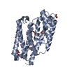
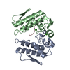
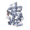
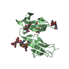
| Title | Crystal structure of bacterial cell-surface alginate-binding protein Algp7 | ||||||
 Components Components | Alginate-binding flagellin | ||||||
 Keywords Keywords | STRUCTURAL PROTEIN / two up-and-down four-helical bundles / alginate binding / alginate | ||||||
| Function / homology |  Function and homology information Function and homology information | ||||||
| Biological species |  Sphingomonas (bacteria) Sphingomonas (bacteria) | ||||||
| Method |  X-RAY DIFFRACTION / SYNCHROTRON / MOLECULAR REPLACEMENT / Resolution: 2.1 Å X-RAY DIFFRACTION / SYNCHROTRON / MOLECULAR REPLACEMENT / Resolution: 2.1 Å | ||||||
 Authors Authors | Maruyama, Y. / Ochiai, A. / Mikami, B. / Hashimoto, W. / Murata, K. | ||||||
 Citation Citation | Journal: Biochem.Biophys.Res.Commun. / Year: 2011 Title: Crystal structure of bacterial cell-surface alginate-binding protein with an M75 peptidase motif. Authors: Maruyama, Y. / Ochiai, A. / Mikami, B. / Hashimoto, W. / Murata, K. | ||||||
| History |
|
- Structure visualization
| Structure viewer | Molecule: MolmilJmol/JSmol |
|---|
- Downloads & links
-Download
| PDBx/mmCIF format | 3at7.cif.gz | 112.2 KB | Display | PDBx/mmCIF format |
|---|---|---|---|---|
| PDB format | pdb3at7.ent.gz | 87.3 KB | Display | PDB format |
| PDBx/mmJSON format | 3at7.json.gz | Tree view | PDBx/mmJSON format | |
| Others |  Other downloads Other downloads |
-Validation report
| Summary document | 3at7_validation.pdf.gz | 430.2 KB | Display | wwPDB validaton report |
|---|---|---|---|---|
| Full document | 3at7_full_validation.pdf.gz | 434.2 KB | Display | |
| Data in XML | 3at7_validation.xml.gz | 21.3 KB | Display | |
| Data in CIF | 3at7_validation.cif.gz | 30.9 KB | Display | |
| Arichive directory | https://data.pdbj.org/pub/pdb/validation_reports/at/3at7ftp://data.pdbj.org/pub/pdb/validation_reports/at/3at7 | HTTPS FTP |
-Related structure data
| Similar structure data |
|---|


-Links
 PDBj
PDBj- Assembly
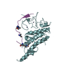
| Deposited unit | 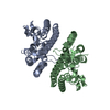
| ||||||||
|---|---|---|---|---|---|---|---|---|---|
| 1 | 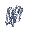
| ||||||||
| 2 | 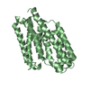
| ||||||||
| Unit cell |
| ||||||||
| Details | AUTHOR DETERMINED BIOLOGICAL UNIT: UNKNOWN |
-Components
| #1: Protein | Mass: 31701.088 Da / Num. of mol.: 2 Source method: isolated from a genetically manipulated source Source: (gene. exp.) Sphingomonas (bacteria) / Strain: A1 / Gene: p7 / Plasmid: pET44a / Production host: #2: Water | ChemComp-HOH / |  Mass: 18.015 Da / Num. of mol.: 267 / Source method: isolated from a natural source / Formula: H2O Mass: 18.015 Da / Num. of mol.: 267 / Source method: isolated from a natural source / Formula: H2O |
|---|
-Experimental details
-Experiment
| Experiment | Method: X-RAY DIFFRACTION / Number of used crystals: 1 |
|---|
- Sample preparation
| Crystal | Density Matthews: 2.12 Å3/Da / Density % sol: 41.88 % |
|---|---|
| Crystal grow | Temperature: 293 K / Method: vapor diffusion / pH: 4.1 Details: 10% PEG 8000, 0.2M NaCl, 0.1M Sodium Citrate pH4.1, VAPOR DIFFUSION, temperature 293K |
-Data collection
| Diffraction | Mean temperature: 100 K |
|---|---|
| Diffraction source | Source: SYNCHROTRON / Site: SPring-8  / Beamline: BL38B1 / Wavelength: 1 Å / Beamline: BL38B1 / Wavelength: 1 Å |
| Detector | Type: ADSC QUANTUM 210 / Detector: CCD / Date: May 23, 2010 |
| Radiation | Protocol: SINGLE WAVELENGTH / Monochromatic (M) / Laue (L): M / Scattering type: x-ray |
| Radiation wavelength | Wavelength: 1 Å / Relative weight: 1 |
| Reflection | Resolution: 2.1→50 Å / Num. obs: 32186 / % possible obs: 99.9 % / Rmerge(I) obs: 0.077 / Net I/σ(I): 34.5 |
| Reflection shell | Resolution: 2.1→2.18 Å / Rmerge(I) obs: 0.305 / % possible all: 100 |
- Processing
| Software |
| ||||||||||||||||||||||||||||||||||||||||||||||||||||||||||||||||||||||||||||||||||||||||||||||||||||||||||||||||||||||||||||||||||||||||||||||||||||||||||||||||||||||||||
|---|---|---|---|---|---|---|---|---|---|---|---|---|---|---|---|---|---|---|---|---|---|---|---|---|---|---|---|---|---|---|---|---|---|---|---|---|---|---|---|---|---|---|---|---|---|---|---|---|---|---|---|---|---|---|---|---|---|---|---|---|---|---|---|---|---|---|---|---|---|---|---|---|---|---|---|---|---|---|---|---|---|---|---|---|---|---|---|---|---|---|---|---|---|---|---|---|---|---|---|---|---|---|---|---|---|---|---|---|---|---|---|---|---|---|---|---|---|---|---|---|---|---|---|---|---|---|---|---|---|---|---|---|---|---|---|---|---|---|---|---|---|---|---|---|---|---|---|---|---|---|---|---|---|---|---|---|---|---|---|---|---|---|---|---|---|---|---|---|---|---|---|
| Refinement | Method to determine structure: MOLECULAR REPLACEMENT / Resolution: 2.1→30 Å / Cor.coef. Fo:Fc: 0.94 / Cor.coef. Fo:Fc free: 0.915 / SU B: 4.529 / SU ML: 0.123 / Cross valid method: THROUGHOUT / ESU R Free: 0.188 / Stereochemistry target values: MAXIMUM LIKELIHOOD / Details: HYDROGENS HAVE BEEN ADDED IN THE RIDING POSITIONS
| ||||||||||||||||||||||||||||||||||||||||||||||||||||||||||||||||||||||||||||||||||||||||||||||||||||||||||||||||||||||||||||||||||||||||||||||||||||||||||||||||||||||||||
| Solvent computation | Ion probe radii: 0.8 Å / Shrinkage radii: 0.8 Å / VDW probe radii: 1.4 Å / Solvent model: MASK | ||||||||||||||||||||||||||||||||||||||||||||||||||||||||||||||||||||||||||||||||||||||||||||||||||||||||||||||||||||||||||||||||||||||||||||||||||||||||||||||||||||||||||
| Displacement parameters | Biso mean: 21.663 Å2
| ||||||||||||||||||||||||||||||||||||||||||||||||||||||||||||||||||||||||||||||||||||||||||||||||||||||||||||||||||||||||||||||||||||||||||||||||||||||||||||||||||||||||||
| Refinement step | Cycle: LAST / Resolution: 2.1→30 Å
| ||||||||||||||||||||||||||||||||||||||||||||||||||||||||||||||||||||||||||||||||||||||||||||||||||||||||||||||||||||||||||||||||||||||||||||||||||||||||||||||||||||||||||
| Refine LS restraints |
| ||||||||||||||||||||||||||||||||||||||||||||||||||||||||||||||||||||||||||||||||||||||||||||||||||||||||||||||||||||||||||||||||||||||||||||||||||||||||||||||||||||||||||
| LS refinement shell | Resolution: 2.1→2.155 Å / Total num. of bins used: 20
|
