 Movie
Movie Controller

[English] 日本語
 Yorodumi
- PDB-3aso: Bovine heart cytochrome C oxidase in the fully oxidized state mea... -
+ Open data

- Basic information
| Entry | Database: PDB / ID: 3aso | ||||||
|---|---|---|---|---|---|---|---|
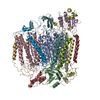
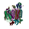
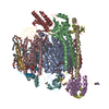
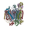
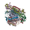
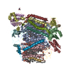
| Title | Bovine heart cytochrome C oxidase in the fully oxidized state measured at 0.9 angstrom wavelength | ||||||
 Components Components | (Cytochrome c oxidase subunit ...) x 13 | ||||||
 Keywords Keywords | OXIDOREDUCTASE | ||||||
| Function / homology |  Function and homology information Function and homology informationComplex IV assembly / TP53 Regulates Metabolic Genes / respiratory chain complex IV assembly / Cytoprotection by HMOX1 / mitochondrial respirasome assembly / Respiratory electron transport / respiratory chain complex IV / respiratory chain complex / Mitochondrial translation termination / cytochrome-c oxidase ...Complex IV assembly / TP53 Regulates Metabolic Genes / respiratory chain complex IV assembly / Cytoprotection by HMOX1 / mitochondrial respirasome assembly / Respiratory electron transport / respiratory chain complex IV / respiratory chain complex / Mitochondrial translation termination / cytochrome-c oxidase / oxidative phosphorylation / mitochondrial electron transport, cytochrome c to oxygen / cytochrome-c oxidase activity / Mitochondrial protein degradation / ATP synthesis coupled electron transport / enzyme regulator activity / proton transmembrane transport / aerobic respiration / central nervous system development / respiratory electron transport chain / mitochondrial membrane / oxidoreductase activity / mitochondrial inner membrane / copper ion binding / heme binding / mitochondrion / metal ion binding Similarity search - Function | ||||||
| Biological species |  | ||||||
| Method |  X-RAY DIFFRACTION / SYNCHROTRON / MOLECULAR REPLACEMENT / Resolution: 2.3 Å X-RAY DIFFRACTION / SYNCHROTRON / MOLECULAR REPLACEMENT / Resolution: 2.3 Å | ||||||
 Authors Authors | Suga, M. / Yano, N. / Muramoto, K. / Shinzawa-Itoh, K. / Maeda, T. / Yamashita, E. / Tsukihara, T. / Yoshikawa, S. | ||||||
 Citation Citation | Journal: Acta Crystallogr.,Sect.D / Year: 2011 Title: Distinguishing between Cl- and O2(2-) as the bridging element between Fe3+ and Cu2+ in resting-oxidized cytochrome c oxidase Authors: Suga, M. / Yano, N. / Muramoto, K. / Shinzawa-Itoh, K. / Maeda, T. / Yamashita, E. / Tsukihara, T. / Yoshikawa, S. | ||||||
| History |
|
- Structure visualization
| Structure viewer | Molecule: MolmilJmol/JSmol |
|---|
- Downloads & links
-Download
| PDBx/mmCIF format | 3aso.cif.gz | 818.7 KB | Display | PDBx/mmCIF format |
|---|---|---|---|---|
| PDB format | pdb3aso.ent.gz | 666.9 KB | Display | PDB format |
| PDBx/mmJSON format | 3aso.json.gz | Tree view | PDBx/mmJSON format | |
| Others |  Other downloads Other downloads |
-Validation report
| Arichive directory | https://data.pdbj.org/pub/pdb/validation_reports/as/3asoftp://data.pdbj.org/pub/pdb/validation_reports/as/3aso | HTTPS FTP |
|---|
-Related structure data
| Related structure data |  3asnC  2dyrS S: Starting model for refinement C: citing same article ( |
|---|---|
| Similar structure data |


-Links
 PDBj
PDBj


- Assembly
| Deposited unit | 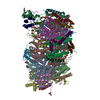
| ||||||||
|---|---|---|---|---|---|---|---|---|---|
| 1 | 
| ||||||||
| 2 | 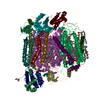
| ||||||||
| Unit cell |
|
-Components
-Cytochrome c oxidase subunit ... , 13 types, 26 molecules ANBOCPDQERFSGTHUIVJWKXLYMZ
| #1: Protein | Mass: 57093.852 Da / Num. of mol.: 2 / Source method: isolated from a natural source / Source: (natural) #2: Protein | Mass: 26068.404 Da / Num. of mol.: 2 / Source method: isolated from a natural source / Source: (natural) #3: Protein | Mass: 29957.627 Da / Num. of mol.: 2 / Source method: isolated from a natural source / Source: (natural) #4: Protein | Mass: 17179.646 Da / Num. of mol.: 2 / Source method: isolated from a natural source / Source: (natural) #5: Protein | Mass: 12453.081 Da / Num. of mol.: 2 / Source method: isolated from a natural source / Source: (natural) #6: Protein | Mass: 10684.038 Da / Num. of mol.: 2 / Source method: isolated from a natural source / Source: (natural) #7: Protein | Mass: 9629.782 Da / Num. of mol.: 2 / Source method: isolated from a natural source / Source: (natural) #8: Protein | Mass: 10039.244 Da / Num. of mol.: 2 / Source method: isolated from a natural source / Source: (natural) #9: Protein | Mass: 8537.019 Da / Num. of mol.: 2 / Source method: isolated from a natural source / Source: (natural) #10: Protein | Mass: 6682.726 Da / Num. of mol.: 2 / Source method: isolated from a natural source / Source: (natural) #11: Protein | Mass: 6365.217 Da / Num. of mol.: 2 / Source method: isolated from a natural source / Source: (natural) #12: Protein/peptide | Mass: 5449.396 Da / Num. of mol.: 2 / Source method: isolated from a natural source / Source: (natural) #13: Protein/peptide | Mass: 4967.756 Da / Num. of mol.: 2 / Source method: isolated from a natural source / Source: (natural) |
|---|
-Sugars , 1 types, 2 molecules 
| #27: Sugar |  Type: D-saccharide / Mass: 482.562 Da / Num. of mol.: 2 Type: D-saccharide / Mass: 482.562 Da / Num. of mol.: 2Source method: isolated from a genetically manipulated source Formula: C22H42O11 / Comment: detergent*YM |
|---|
-Non-polymers , 14 types, 1761 molecules 


| #14: Chemical | ChemComp-HEA /  Mass: 852.837 Da / Num. of mol.: 4 / Source method: obtained synthetically / Formula: C49H56FeN4O6 Mass: 852.837 Da / Num. of mol.: 4 / Source method: obtained synthetically / Formula: C49H56FeN4O6#15: Chemical |  Mass: 63.546 Da / Num. of mol.: 2 / Source method: obtained synthetically / Formula: Cu Mass: 63.546 Da / Num. of mol.: 2 / Source method: obtained synthetically / Formula: Cu#16: Chemical |  Mass: 24.305 Da / Num. of mol.: 2 / Source method: obtained synthetically / Formula: Mg Mass: 24.305 Da / Num. of mol.: 2 / Source method: obtained synthetically / Formula: Mg#17: Chemical |  Mass: 22.990 Da / Num. of mol.: 2 / Source method: obtained synthetically / Formula: Na Mass: 22.990 Da / Num. of mol.: 2 / Source method: obtained synthetically / Formula: Na#18: Chemical | ChemComp-TGL /  Mass: 891.480 Da / Num. of mol.: 6 / Source method: obtained synthetically / Formula: C57H110O6 Mass: 891.480 Da / Num. of mol.: 6 / Source method: obtained synthetically / Formula: C57H110O6#19: Chemical | ChemComp-PGV / (  Mass: 749.007 Da / Num. of mol.: 8 / Source method: obtained synthetically / Formula: C40H77O10P / Comment: phospholipid*YM Mass: 749.007 Da / Num. of mol.: 8 / Source method: obtained synthetically / Formula: C40H77O10P / Comment: phospholipid*YM#20: Chemical |  Mass: 127.092 Da / Num. of mol.: 2 / Source method: obtained synthetically / Formula: Cu2 Mass: 127.092 Da / Num. of mol.: 2 / Source method: obtained synthetically / Formula: Cu2#21: Chemical |  Mass: 759.068 Da / Num. of mol.: 2 / Source method: obtained synthetically / Formula: C42H81NO8P / Comment: phospholipid*YM Mass: 759.068 Da / Num. of mol.: 2 / Source method: obtained synthetically / Formula: C42H81NO8P / Comment: phospholipid*YM#22: Chemical | ChemComp-CHD /  Mass: 408.571 Da / Num. of mol.: 8 / Source method: obtained synthetically / Formula: C24H40O5 Mass: 408.571 Da / Num. of mol.: 8 / Source method: obtained synthetically / Formula: C24H40O5#23: Chemical |  Num. of mol.: 2 / Source method: obtained synthetically Num. of mol.: 2 / Source method: obtained synthetically#24: Chemical | ChemComp-PEK / (  Mass: 768.055 Da / Num. of mol.: 6 / Source method: obtained synthetically / Formula: C43H78NO8P / Comment: phospholipid*YM Mass: 768.055 Da / Num. of mol.: 6 / Source method: obtained synthetically / Formula: C43H78NO8P / Comment: phospholipid*YM#25: Chemical | ChemComp-CDL /  Mass: 1464.043 Da / Num. of mol.: 4 / Source method: obtained synthetically / Formula: C81H156O17P2 / Comment: phospholipid*YM Mass: 1464.043 Da / Num. of mol.: 4 / Source method: obtained synthetically / Formula: C81H156O17P2 / Comment: phospholipid*YM#26: Chemical |  Mass: 65.409 Da / Num. of mol.: 2 / Source method: obtained synthetically / Formula: Zn Mass: 65.409 Da / Num. of mol.: 2 / Source method: obtained synthetically / Formula: Zn#28: Water | ChemComp-HOH / | Mass: 18.015 Da / Num. of mol.: 1711 / Source method: isolated from a natural source / Formula: H2O |
|---|
-Experimental details
-Experiment
| Experiment | Method: X-RAY DIFFRACTION / Number of used crystals: 1 |
|---|
- Sample preparation
| Crystal | Density Matthews: 4.02 Å3/Da / Density % sol: 69.43 % |
|---|---|
| Crystal grow | Temperature: 277 K / Method: batch / pH: 6.8 Details: 40mM sodium phosphate, 0.2% decylmaltoside , pH 6.8, BATCH, temperature 277K |
-Data collection
| Diffraction | Mean temperature: 50 K |
|---|---|
| Diffraction source | Source: SYNCHROTRON / Site: SPring-8  / Beamline: BL44XU / Wavelength: 0.9 Å / Beamline: BL44XU / Wavelength: 0.9 Å |
| Detector | Type: RAYONIX MX225HE / Detector: CCD / Date: Jun 14, 2009 |
| Radiation | Protocol: SINGLE WAVELENGTH / Monochromatic (M) / Laue (L): M / Scattering type: x-ray |
| Radiation wavelength | Wavelength: 0.9 Å / Relative weight: 1 |
| Reflection | Resolution: 2.3→108 Å / Num. obs: 286689 / % possible obs: 99.9 % / Observed criterion σ(I): -3 |
| Reflection shell | Resolution: 2.3→2.33 Å / % possible all: 99.9 |
- Processing
| Software |
| ||||||||||||||||||
|---|---|---|---|---|---|---|---|---|---|---|---|---|---|---|---|---|---|---|---|
| Refinement | Method to determine structure: MOLECULAR REPLACEMENT Starting model: 2DYR Resolution: 2.3→15 Å
| ||||||||||||||||||
| Refinement step | Cycle: LAST / Resolution: 2.3→15 Å
| ||||||||||||||||||
| Refine LS restraints |
|
