 Movie
Movie Controller


+ Open data

- Basic information
| Entry | Database: PDB / ID: 3aqs | ||||||
|---|---|---|---|---|---|---|---|
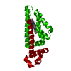
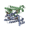
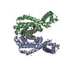
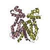
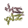
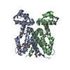
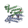
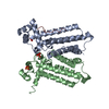
| Title | Crystal structure of RolR (NCGL1110) without ligand | ||||||
 Components Components | Bacterial regulatory proteins, tetR family | ||||||
 Keywords Keywords | TRANSCRIPTION REGULATOR / HELIX-TURN-HELIX / ALL ALPHA / TRANSCRIPTION / TRANSCRIPTION REGULATION / all helix / TetR family / transcriptional repressor / resorcinol binding / DNA binding | ||||||
| Function / homology |  Function and homology information Function and homology information | ||||||
| Biological species |  Corynebacterium glutamicum (bacteria) Corynebacterium glutamicum (bacteria) | ||||||
| Method |  X-RAY DIFFRACTION / SYNCHROTRON / MOLECULAR REPLACEMENT / Resolution: 3.6 Å X-RAY DIFFRACTION / SYNCHROTRON / MOLECULAR REPLACEMENT / Resolution: 3.6 Å | ||||||
 Authors Authors | Li, D.F. / Zhang, N. / Hou, Y.J. / Liu, S.J. / Wang, D.C. | ||||||
 Citation Citation | Journal: Plos One / Year: 2011 Title: Crystal structures of the transcriptional repressor RolR reveals a novel recognition mechanism between inducer and regulator. Authors: Li, D.F. / Zhang, N. / Hou, Y.J. / Huang, Y. / Hu, Y. / Zhang, Y. / Liu, S.J. / Wang, D.C. | ||||||
| History |
|
- Structure visualization
| Structure viewer | Molecule: MolmilJmol/JSmol |
|---|
- Downloads & links
-Download
| PDBx/mmCIF format | 3aqs.cif.gz | 138.5 KB | Display | PDBx/mmCIF format |
|---|---|---|---|---|
| PDB format | pdb3aqs.ent.gz | 111.3 KB | Display | PDB format |
| PDBx/mmJSON format | 3aqs.json.gz | Tree view | PDBx/mmJSON format | |
| Others |  Other downloads Other downloads |
-Validation report
| Arichive directory | https://data.pdbj.org/pub/pdb/validation_reports/aq/3aqsftp://data.pdbj.org/pub/pdb/validation_reports/aq/3aqs | HTTPS FTP |
|---|
-Related structure data


-Links
 PDBj
PDBj- Assembly
| Deposited unit | 
| ||||||||
|---|---|---|---|---|---|---|---|---|---|
| 1 | 
| ||||||||
| 2 | 
| ||||||||
| Unit cell |
|
-Components
| #1: Protein | Mass: 26905.332 Da / Num. of mol.: 4 Source method: isolated from a genetically manipulated source Source: (gene. exp.) Corynebacterium glutamicum (bacteria) / Strain: ATCC 13032 / Gene: cg1308, Cgl1157, ncgl1110 / Plasmid: PET28A / Production host: |
|---|
-Experimental details
-Experiment
| Experiment | Method: X-RAY DIFFRACTION / Number of used crystals: 1 |
|---|
- Sample preparation
| Crystal | Density Matthews: 3.19 Å3/Da / Density % sol: 61.47 % |
|---|---|
| Crystal grow | Temperature: 300 K / Method: vapor diffusion, hanging drop / pH: 8 Details: 3M ammonium sulfate, pH 8.0, VAPOR DIFFUSION, HANGING DROP, temperature 300K |
-Data collection
| Diffraction | Mean temperature: 90 K |
|---|---|
| Diffraction source | Source: SYNCHROTRON / Site: Photon Factory  / Beamline: BL-5A / Wavelength: 1 Å / Beamline: BL-5A / Wavelength: 1 Å |
| Detector | Type: ADSC QUANTUM 315 / Detector: CCD / Date: Jun 18, 2007 |
| Radiation | Monochromator: Numerical link type Si(111) double crystal monochromator Protocol: SINGLE WAVELENGTH / Monochromatic (M) / Laue (L): M / Scattering type: x-ray |
| Radiation wavelength | Wavelength: 1 Å / Relative weight: 1 |
| Reflection | Resolution: 3.6→108.46 Å / Num. all: 16716 / Num. obs: 16353 / % possible obs: 98.1 % / Observed criterion σ(F): 0 / Observed criterion σ(I): 0 / Redundancy: 5.7 % / Biso Wilson estimate: 45.4 Å2 / Rmerge(I) obs: 0.166 / Rsym value: 0.166 / Net I/σ(I): 13 |
| Reflection shell | Resolution: 3.6→3.79 Å / Redundancy: 5 % / Rmerge(I) obs: 0.451 / Mean I/σ(I) obs: 2.9 / Num. unique all: 2238 / Rsym value: 0.451 / % possible all: 93.8 |
- Processing
| Software |
| ||||||||||||||||||||||||||||||||||||||||||||||||||||||||||||
|---|---|---|---|---|---|---|---|---|---|---|---|---|---|---|---|---|---|---|---|---|---|---|---|---|---|---|---|---|---|---|---|---|---|---|---|---|---|---|---|---|---|---|---|---|---|---|---|---|---|---|---|---|---|---|---|---|---|---|---|---|---|
| Refinement | Method to determine structure: MOLECULAR REPLACEMENT / Resolution: 3.6→93.17 Å / Rfactor Rfree error: 0.007 / Data cutoff high absF: 2239647.52 / Data cutoff low absF: 0 / Isotropic thermal model: RESTRAINED / Cross valid method: THROUGHOUT / σ(F): 0 / σ(I): 0 / Stereochemistry target values: Engh & Huber / Details: BULK SOLVENT MODEL USED
| ||||||||||||||||||||||||||||||||||||||||||||||||||||||||||||
| Solvent computation | Solvent model: FLAT MODEL / Bsol: 75.8428 Å2 / ksol: 0.35 e/Å3 | ||||||||||||||||||||||||||||||||||||||||||||||||||||||||||||
| Displacement parameters | Biso mean: 103 Å2
| ||||||||||||||||||||||||||||||||||||||||||||||||||||||||||||
| Refine analyze |
| ||||||||||||||||||||||||||||||||||||||||||||||||||||||||||||
| Refinement step | Cycle: LAST / Resolution: 3.6→93.17 Å
| ||||||||||||||||||||||||||||||||||||||||||||||||||||||||||||
| Refine LS restraints |
| ||||||||||||||||||||||||||||||||||||||||||||||||||||||||||||
| LS refinement shell | Resolution: 3.6→3.83 Å / Rfactor Rfree error: 0.025 / Total num. of bins used: 6
| ||||||||||||||||||||||||||||||||||||||||||||||||||||||||||||
| Xplor file |
|
