 Movie
Movie Controller


+ Open data

- Basic information
| Entry | Database: PDB / ID: 3aqm | ||||||
|---|---|---|---|---|---|---|---|
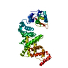
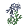
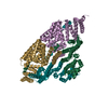
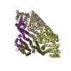
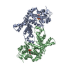
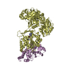
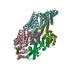
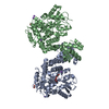
| Title | Structure of bacterial protein (form II) | ||||||
 Components Components | Poly(A) polymerase | ||||||
 Keywords Keywords | TRANSFERASE / TRANSFERASE/RNA / ATP-BINDING / NUCLEOTIDE-BINDING / RNA-BINDING / NUCLEOTIDYLTRANSFERASE / ATP Binding / A-Phosphorylation | ||||||
| Function / homology | cca-adding enzyme, domain 2 / cca-adding enzyme, domain 2 / Beta Polymerase, domain 2 / Beta Polymerase; domain 2 / 2-Layer Sandwich / Orthogonal Bundle / Mainly Alpha / Alpha Beta / :  Function and homology information Function and homology information | ||||||
| Biological species |  | ||||||
| Method |  X-RAY DIFFRACTION / SYNCHROTRON / MOLECULAR REPLACEMENT / Resolution: 3.15 Å X-RAY DIFFRACTION / SYNCHROTRON / MOLECULAR REPLACEMENT / Resolution: 3.15 Å | ||||||
 Authors Authors | Toh, Y. / Takeshita, D. / Tomita , K. | ||||||
 Citation Citation | Journal: Structure / Year: 2011 Title: Mechanism for the alteration of the substrate specificities of template-independent RNA polymerases Authors: Toh, Y. / Takeshita, D. / Nagaike, T. / Numata, T. / Tomita, K. | ||||||
| History |
|
- Structure visualization
| Structure viewer | Molecule: MolmilJmol/JSmol |
|---|
- Downloads & links
-Download
| PDBx/mmCIF format | 3aqm.cif.gz | 166.7 KB | Display | PDBx/mmCIF format |
|---|---|---|---|---|
| PDB format | pdb3aqm.ent.gz | 132.5 KB | Display | PDB format |
| PDBx/mmJSON format | 3aqm.json.gz | Tree view | PDBx/mmJSON format | |
| Others |  Other downloads Other downloads |
-Validation report
| Arichive directory | https://data.pdbj.org/pub/pdb/validation_reports/aq/3aqmftp://data.pdbj.org/pub/pdb/validation_reports/aq/3aqm | HTTPS FTP |
|---|
-Related structure data


-Links
 PDBj
PDBj- Assembly
| Deposited unit | 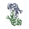
| ||||||||
|---|---|---|---|---|---|---|---|---|---|
| 1 |
| ||||||||
| Unit cell |
|
-Components
| #1: Protein | Mass: 48034.160 Da / Num. of mol.: 2 / Fragment: UNP RESIDUES 17-431 Source method: isolated from a genetically manipulated source Source: (gene. exp.) References: UniProt: C9QS13, polynucleotide adenylyltransferase #2: Chemical |   Mass: 24.305 Da / Num. of mol.: 2 / Source method: obtained synthetically / Formula: Mg Mass: 24.305 Da / Num. of mol.: 2 / Source method: obtained synthetically / Formula: Mg |
|---|
-Experimental details
-Experiment
| Experiment | Method: X-RAY DIFFRACTION / Number of used crystals: 1 |
|---|
- Sample preparation
| Crystal | Density Matthews: 4.08 Å3/Da / Density % sol: 69.86 % |
|---|---|
| Crystal grow | Temperature: 293 K / Method: vapor diffusion, hanging drop / pH: 8 Details: 100mM Tris-Cl, pH 8.0, 1.2 M sodium acetate, 5mM MgCl2, VAPOR DIFFUSION, HANGING DROP, temperature 293K |
-Data collection
| Diffraction | Mean temperature: 100 K |
|---|---|
| Diffraction source | Source: SYNCHROTRON / Site: Photon Factory  / Beamline: BL-17A / Wavelength: 1 Å / Beamline: BL-17A / Wavelength: 1 Å |
| Detector | Type: ADSC QUANTUM 270 / Detector: CCD / Date: Oct 3, 2010 |
| Radiation | Monochromator: SI(111) / Protocol: SINGLE WAVELENGTH / Monochromatic (M) / Laue (L): M / Scattering type: x-ray |
| Radiation wavelength | Wavelength: 1 Å / Relative weight: 1 |
| Reflection | Resolution: 3.15→50 Å / Num. obs: 28023 / % possible obs: 99.5 % / Observed criterion σ(F): 2.1 / Observed criterion σ(I): 14.5 |
| Reflection shell | Resolution: 3.15→3.26 Å / Redundancy: 5.6 % / Rmerge(I) obs: 0.462 / Mean I/σ(I) obs: 2.1 / Num. unique all: 28023 / % possible all: 99.7 |
- Processing
| Software |
| ||||||||||||||||||||||||||||||||||||||||||||||||||||||||||||||||||||||||||||||||
|---|---|---|---|---|---|---|---|---|---|---|---|---|---|---|---|---|---|---|---|---|---|---|---|---|---|---|---|---|---|---|---|---|---|---|---|---|---|---|---|---|---|---|---|---|---|---|---|---|---|---|---|---|---|---|---|---|---|---|---|---|---|---|---|---|---|---|---|---|---|---|---|---|---|---|---|---|---|---|---|---|---|
| Refinement | Method to determine structure: MOLECULAR REPLACEMENT / Resolution: 3.15→29.8 Å / Rfactor Rfree error: 0.008 / Data cutoff high absF: 8691641.76 / Data cutoff low absF: 0 / Isotropic thermal model: RESTRAINED / Cross valid method: THROUGHOUT / σ(F): 0 / Stereochemistry target values: Engh & Huber / Details: BULK SOLVENT MODEL USED
| ||||||||||||||||||||||||||||||||||||||||||||||||||||||||||||||||||||||||||||||||
| Solvent computation | Solvent model: FLAT MODEL / Bsol: 23.2056 Å2 / ksol: 0.28 e/Å3 | ||||||||||||||||||||||||||||||||||||||||||||||||||||||||||||||||||||||||||||||||
| Displacement parameters | Biso mean: 105.4 Å2
| ||||||||||||||||||||||||||||||||||||||||||||||||||||||||||||||||||||||||||||||||
| Refine analyze |
| ||||||||||||||||||||||||||||||||||||||||||||||||||||||||||||||||||||||||||||||||
| Refinement step | Cycle: LAST / Resolution: 3.15→29.8 Å
| ||||||||||||||||||||||||||||||||||||||||||||||||||||||||||||||||||||||||||||||||
| Refine LS restraints |
| ||||||||||||||||||||||||||||||||||||||||||||||||||||||||||||||||||||||||||||||||
| LS refinement shell | Resolution: 3.15→3.35 Å / Rfactor Rfree error: 0.036 / Total num. of bins used: 6
| ||||||||||||||||||||||||||||||||||||||||||||||||||||||||||||||||||||||||||||||||
| Xplor file |
|
