 Movie
Movie Controller

[English] 日本語
 Yorodumi
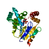
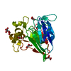
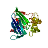
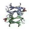
- PDB-3akd: Crystal structure of CMP kinase in complex with CDP from Thermus ... -
+ Open data

- Basic information
| Entry | Database: PDB / ID: 3akd | ||||||
|---|---|---|---|---|---|---|---|
| Title | Crystal structure of CMP kinase in complex with CDP from Thermus thermophilus HB8 | ||||||
 Components Components | Cytidylate kinase | ||||||
 Keywords Keywords | TRANSFERASE / CMP kinase / CDP complex / open conformation / nucleotide metabolism | ||||||
| Function / homology |  Function and homology information Function and homology information(d)CMP kinase / CMP kinase activity / dCMP kinase activity / pyrimidine nucleotide metabolic process / ATP binding / cytoplasm Similarity search - Function | ||||||
| Biological species |   Thermus thermophilus (bacteria) Thermus thermophilus (bacteria) | ||||||
| Method |  X-RAY DIFFRACTION / SYNCHROTRON / MOLECULAR REPLACEMENT / Resolution: 1.6 Å X-RAY DIFFRACTION / SYNCHROTRON / MOLECULAR REPLACEMENT / Resolution: 1.6 Å | ||||||
 Authors Authors | Mega, R. / Nakagawa, N. / Kuramitsu, S. / Masui, R. | ||||||
 Citation Citation | Journal: To be Published Title: The crystal structure of the tertiary complex of CMP kinase with a phosphoryl group acceptor and a donor from Thermus thermophilus HB8 Authors: Mega, R. / Nakagawa, N. / Kuramitsu, S. / Masui, R. | ||||||
| History |
|
- Structure visualization
| Structure viewer | Molecule: MolmilJmol/JSmol |
|---|
- Downloads & links
-Download
| PDBx/mmCIF format | 3akd.cif.gz | 54.6 KB | Display | PDBx/mmCIF format |
|---|---|---|---|---|
| PDB format | pdb3akd.ent.gz | 38.5 KB | Display | PDB format |
| PDBx/mmJSON format | 3akd.json.gz | Tree view | PDBx/mmJSON format | |
| Others |  Other downloads Other downloads |
-Validation report
| Summary document | 3akd_validation.pdf.gz | 804.7 KB | Display | wwPDB validaton report |
|---|---|---|---|---|
| Full document | 3akd_full_validation.pdf.gz | 809.4 KB | Display | |
| Data in XML | 3akd_validation.xml.gz | 11.7 KB | Display | |
| Data in CIF | 3akd_validation.cif.gz | 15.8 KB | Display | |
| Arichive directory | https://data.pdbj.org/pub/pdb/validation_reports/ak/3akdftp://data.pdbj.org/pub/pdb/validation_reports/ak/3akd | HTTPS FTP |
-Related structure data
| Related structure data | 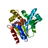 3akcC 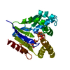 2cmkS C: citing same article ( S: Starting model for refinement |
|---|---|
| Similar structure data |


-Links
 PDBj
PDBj- Assembly
| Deposited unit | 
| ||||||||
|---|---|---|---|---|---|---|---|---|---|
| 1 |
| ||||||||
| Unit cell |
|
-Components
| #1: Protein | Mass: 22584.818 Da / Num. of mol.: 1 Source method: isolated from a genetically manipulated source Source: (gene. exp.) Thermus thermophilus (bacteria) / Strain: HB8 / Gene: TTHA0458 / Plasmid: pET-11a / Production host: |
|---|---|
| #2: Chemical | ChemComp-CDP / 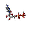  Mass: 403.176 Da / Num. of mol.: 1 / Source method: obtained synthetically / Formula: C9H15N3O11P2 Mass: 403.176 Da / Num. of mol.: 1 / Source method: obtained synthetically / Formula: C9H15N3O11P2 |
| #3: Water | ChemComp-HOH /  Mass: 18.015 Da / Num. of mol.: 103 / Source method: isolated from a natural source / Formula: H2O Mass: 18.015 Da / Num. of mol.: 103 / Source method: isolated from a natural source / Formula: H2O |
-Experimental details
-Experiment
| Experiment | Method: X-RAY DIFFRACTION / Number of used crystals: 1 |
|---|
- Sample preparation
| Crystal | Density Matthews: 2.72 Å3/Da / Density % sol: 54.83 % |
|---|---|
| Crystal grow | Temperature: 293 K / Method: vapor diffusion, sitting drop / pH: 7 Details: 0.1M BIS-TRIS propane, 3.5M sodium formate, pH 7.0, VAPOR DIFFUSION, SITTING DROP, temperature 293K |
-Data collection
| Diffraction | Mean temperature: 90 K |
|---|---|
| Diffraction source | Source: SYNCHROTRON / Site: SPring-8  / Beamline: BL45XU / Wavelength: 1 Å / Beamline: BL45XU / Wavelength: 1 Å |
| Detector | Type: ADSC QUANTUM 210 / Detector: CCD / Date: Jun 6, 2006 |
| Radiation | Monochromator: transparent diamond double crystal / Protocol: SINGLE WAVELENGTH / Monochromatic (M) / Laue (L): M / Scattering type: x-ray |
| Radiation wavelength | Wavelength: 1 Å / Relative weight: 1 |
| Reflection | Resolution: 1.6→50 Å / Num. obs: 29707 / % possible obs: 93.1 % / Observed criterion σ(I): -3 / Redundancy: 11.1 % / Biso Wilson estimate: 19.9 Å2 / Rmerge(I) obs: 0.038 / Net I/σ(I): 53 |
| Reflection shell | Resolution: 1.6→1.66 Å / Redundancy: 7.7 % / Rmerge(I) obs: 0.262 / Mean I/σ(I) obs: 3.4 / Num. unique all: 1768 / % possible all: 55.7 |
- Processing
| Software |
| |||||||||||||||||||||||||
|---|---|---|---|---|---|---|---|---|---|---|---|---|---|---|---|---|---|---|---|---|---|---|---|---|---|---|
| Refinement | Method to determine structure: MOLECULAR REPLACEMENT Starting model: PDB ENTRY 2CMK Resolution: 1.6→38.58 Å / Rfactor Rfree error: 0.004 / Data cutoff high absF: 1453360.21 / Data cutoff low absF: 0 / Isotropic thermal model: RESTRAINED / Cross valid method: THROUGHOUT / σ(F): 0 / Stereochemistry target values: Engh & Huber / Details: BULK SOLVENT MODEL USED
| |||||||||||||||||||||||||
| Solvent computation | Solvent model: FLAT MODEL / Bsol: 43.9677 Å2 / ksol: 0.4 e/Å3 | |||||||||||||||||||||||||
| Displacement parameters | Biso mean: 22.3 Å2
| |||||||||||||||||||||||||
| Refine analyze |
| |||||||||||||||||||||||||
| Refinement step | Cycle: LAST / Resolution: 1.6→38.58 Å
| |||||||||||||||||||||||||
| Refine LS restraints |
| |||||||||||||||||||||||||
| LS refinement shell | Resolution: 1.6→1.7 Å / Rfactor Rfree error: 0.016
| |||||||||||||||||||||||||
| Xplor file |
|
