- PDB-3aih: Human OS-9 MRH domain complexed with alpha3,alpha6-Man5 -
+
Open data
ID or keywords:
Loading...
-
Basic information
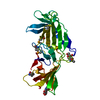
Entry
Database: PDB / ID: 3aih
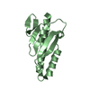
Title
Human OS-9 MRH domain complexed with alpha3,alpha6-Man5
Components
Protein OS-9
Keywords
SUGAR BINDING PROTEIN / Beta barrel / Lectin
Function / homology
Function and homology information
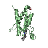
glycosylation-dependent protein binding / negative regulation of retrograde protein transport, ER to cytosol / Hrd1p ubiquitin ligase complex / protein retention in ER lumen / retrograde protein transport, ER to cytosol / AMPK-induced ERAD and lysosome mediated degradation of PD-L1(CD274) / protein targeting / endoplasmic reticulum unfolded protein response / ERAD pathway / ER Quality Control Compartment (ERQC) ...glycosylation-dependent protein binding / negative regulation of retrograde protein transport, ER to cytosol / Hrd1p ubiquitin ligase complex / protein retention in ER lumen / retrograde protein transport, ER to cytosol / AMPK-induced ERAD and lysosome mediated degradation of PD-L1(CD274) / protein targeting / endoplasmic reticulum unfolded protein response / ERAD pathway / ER Quality Control Compartment (ERQC) / response to endoplasmic reticulum stress / Hh mutants are degraded by ERAD / Hedgehog ligand biogenesis / Defective CFTR causes cystic fibrosis / ABC-family protein mediated transport / carbohydrate binding / protease binding / ubiquitin-dependent protein catabolic process / protein ubiquitination / endoplasmic reticulum lumen / endoplasmic reticulum membrane / endoplasmic reticulum Similarity search - Function
Protein OS9-like domain / Protein OS-9-like / Glucosidase II beta subunit-like protein / Cation-dependent Mannose-6-phosphate Receptor; Chain A / Mannose-6-phosphate receptor binding domain / Mannose-6-phosphate receptor binding domain superfamily / MRH domain / MRH domain profile. / Distorted Sandwich / Mainly Beta Similarity search - Domain/homology
Monochromator: Si(111) / Protocol: SINGLE WAVELENGTH / Monochromatic (M) / Laue (L): M / Scattering type: x-ray
Radiation wavelength
Wavelength: 1 Å / Relative weight: 1
Reflection
Resolution: 2.1→50 Å / Num. all: 17542 / Num. obs: 17480 / % possible obs: 99.7 % / Redundancy: 11.4 % / Biso Wilson estimate: 30.3 Å2 / Rmerge(I) obs: 0.098 / Net I/σ(I): 28.5
Reflection shell
Resolution: 2.1→2.18 Å / Redundancy: 11.7 % / Rmerge(I) obs: 0.469 / Mean I/σ(I) obs: 5.5 / Num. unique all: 1694 / % possible all: 100
-
Processing
Software
Name
Version
Classification
HKL-2000
datacollection
SHELXS
phasing
REFMAC
5.5.0102
refinement
DENZO
datareduction
SCALEPACK
datascaling
Refinement
Method to determine structure: SAD / Resolution: 2.1→40 Å / Cor.coef. Fo:Fc: 0.934 / Cor.coef. Fo:Fc free: 0.911 / SU B: 5.571 / SU ML: 0.147 / Cross valid method: THROUGHOUT / ESU R Free: 0.204 / Stereochemistry target values: MAXIMUM LIKELIHOOD / Details: HYDROGENS HAVE BEEN ADDED IN THE RIDING POSITIONS
Rfactor
Num. reflection
% reflection
Selection details
Rfree
0.277
881
5.1 %
RANDOM
Rwork
0.228
-
-
-
obs
0.23
16555
99.89 %
-
all
-
16555
-
-
Solvent computation
Ion probe radii: 0.8 Å / Shrinkage radii: 0.8 Å / VDW probe radii: 1.4 Å / Solvent model: MASK
Displacement parameters
Biso mean: 31.263 Å2
Baniso -1
Baniso -2
Baniso -3
1-
-1.19 Å2
-0.59 Å2
0 Å2
2-
-
-1.19 Å2
0 Å2
3-
-
-
1.78 Å2
Refinement step
Cycle: LAST / Resolution: 2.1→40 Å
Protein
Nucleic acid
Ligand
Solvent
Total
Num. atoms
1836
0
68
79
1983
Refine LS restraints
Refine-ID
Type
Dev ideal
Dev ideal target
Number
X-RAY DIFFRACTION
r_bond_refined_d
0.018
0.021
1939
X-RAY DIFFRACTION
r_angle_refined_deg
1.663
1.968
2626
X-RAY DIFFRACTION
r_dihedral_angle_1_deg
7.276
5
216
X-RAY DIFFRACTION
r_dihedral_angle_2_deg
31.427
22.981
104
X-RAY DIFFRACTION
r_dihedral_angle_3_deg
18.231
15
310
X-RAY DIFFRACTION
r_dihedral_angle_4_deg
17.291
15
17
X-RAY DIFFRACTION
r_chiral_restr
0.123
0.2
271
X-RAY DIFFRACTION
r_gen_planes_refined
0.008
0.021
1473
X-RAY DIFFRACTION
r_mcbond_it
1.133
1.5
1092
X-RAY DIFFRACTION
r_mcangle_it
2.103
2
1749
X-RAY DIFFRACTION
r_scbond_it
2.774
3
847
X-RAY DIFFRACTION
r_scangle_it
4.405
4.5
877
LS refinement shell
Resolution: 2.1→2.154 Å / Total num. of bins used: 20
Rfactor
Num. reflection
% reflection
Rfree
0.317
54
-
Rwork
0.267
1205
-
obs
-
-
100 %
+
About Yorodumi
-
News
-
Feb 9, 2022. New format data for meta-information of EMDB entries
New format data for meta-information of EMDB entries
Version 3 of the EMDB header file is now the official format.
The previous official version 1.9 will be removed from the archive.
In the structure databanks used in Yorodumi, some data are registered as the other names, "COVID-19 virus" and "2019-nCoV". Here are the details of the virus and the list of structure data.
Jan 31, 2019. EMDB accession codes are about to change! (news from PDBe EMDB page)
EMDB accession codes are about to change! (news from PDBe EMDB page)
The allocation of 4 digits for EMDB accession codes will soon come to an end. Whilst these codes will remain in use, new EMDB accession codes will include an additional digit and will expand incrementally as the available range of codes is exhausted. The current 4-digit format prefixed with “EMD-” (i.e. EMD-XXXX) will advance to a 5-digit format (i.e. EMD-XXXXX), and so on. It is currently estimated that the 4-digit codes will be depleted around Spring 2019, at which point the 5-digit format will come into force.
The EM Navigator/Yorodumi systems omit the EMD- prefix.
Related info.:Q: What is EMD? / ID/Accession-code notation in Yorodumi/EM Navigator
Yorodumi is a browser for structure data from EMDB, PDB, SASBDB, etc.
This page is also the successor to EM Navigator detail page, and also detail information page/front-end page for Omokage search.
The word "yorodu" (or yorozu) is an old Japanese word meaning "ten thousand". "mi" (miru) is to see.
Related info.:EMDB / PDB / SASBDB / Comparison of 3 databanks / Yorodumi Search / Aug 31, 2016. New EM Navigator & Yorodumi / Yorodumi Papers / Jmol/JSmol / Function and homology information / Changes in new EM Navigator and Yorodumi
 Movie
Movie Controller


 Open data
 Basic information
 Components
 Keywords
 Function and homology information
 Homo sapiens (human)
 X-RAY DIFFRACTION /
  Authors
 Citation
 Structure visualization
 Downloads & links
 Other downloads
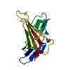


 PDBj
PDBj

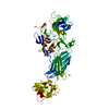
 Assembly


 Mass: 18.015 Da / Num. of mol.: 79 / Source method: isolated from a natural source / Formula: H2O
 Sample preparation
 / Beamline: BL13B1 / Wavelength: 1 Å
 Processing