 Movie
Movie Controller

[English] 日本語
 Yorodumi
- PDB-3ai3: The crystal structure of L-Sorbose reductase from Gluconobacter f... -
+ Open data

- Basic information
| Entry | Database: PDB / ID: 3ai3 | ||||||
|---|---|---|---|---|---|---|---|
| Title | The crystal structure of L-Sorbose reductase from Gluconobacter frateurii complexed with NADPH and L-sorbose | ||||||
 Components Components | NADPH-sorbose reductase | ||||||
 Keywords Keywords | OXIDOREDUCTASE / Rossmann-fold / NADPH-dependent reductase / short chain dehydrogenase/reductase | ||||||
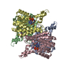
| Function / homology |  Function and homology information Function and homology information | ||||||
| Biological species |  Gluconobacter frateurii (bacteria) Gluconobacter frateurii (bacteria) | ||||||
| Method |  X-RAY DIFFRACTION / SYNCHROTRON / MOLECULAR REPLACEMENT / Resolution: 1.8 Å X-RAY DIFFRACTION / SYNCHROTRON / MOLECULAR REPLACEMENT / Resolution: 1.8 Å | ||||||
 Authors Authors | Kubota, K. / Nagata, K. / Okai, M. / Miyazono, K. / Tanokura, M. | ||||||
 Citation Citation | Journal: J.Mol.Biol. / Year: 2011 Title: The Crystal Structure of l-Sorbose Reductase from Gluconobacter frateurii Complexed with NADPH and l-Sorbose Authors: Kubota, K. / Nagata, K. / Okai, M. / Miyazono, K. / Soemphol, W. / Ohtsuka, J. / Yamamura, A. / Saichana, N. / Toyama, H. / Matsushita, K. / Tanokura, M. | ||||||
| History |
|
- Structure visualization
| Structure viewer | Molecule: MolmilJmol/JSmol |
|---|
- Downloads & links
-Download
| PDBx/mmCIF format | 3ai3.cif.gz | 213.9 KB | Display | PDBx/mmCIF format |
|---|---|---|---|---|
| PDB format | pdb3ai3.ent.gz | 174 KB | Display | PDB format |
| PDBx/mmJSON format | 3ai3.json.gz | Tree view | PDBx/mmJSON format | |
| Others |  Other downloads Other downloads |
-Validation report
| Arichive directory | https://data.pdbj.org/pub/pdb/validation_reports/ai/3ai3ftp://data.pdbj.org/pub/pdb/validation_reports/ai/3ai3 | HTTPS FTP |
|---|
-Related structure data
| Related structure data | 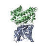 3ai1C  3ai2SC C: citing same article ( S: Starting model for refinement |
|---|---|
| Similar structure data |


-Links
 PDBj
PDBj


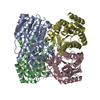
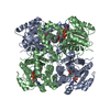
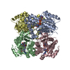
- Assembly
| Deposited unit | 
| ||||||||
|---|---|---|---|---|---|---|---|---|---|
| 1 |
| ||||||||
| Unit cell |
|
-Components
| #1: Protein | Mass: 28326.541 Da / Num. of mol.: 4 / Mutation: H116L Source method: isolated from a genetically manipulated source Source: (gene. exp.) Gluconobacter frateurii (bacteria) / Gene: sboA / Plasmid: pET28a / Production host: #2: Chemical | ChemComp-NDP /   Mass: 745.421 Da / Num. of mol.: 4 / Source method: obtained synthetically / Formula: C21H30N7O17P3 Mass: 745.421 Da / Num. of mol.: 4 / Source method: obtained synthetically / Formula: C21H30N7O17P3#3: Sugar | ChemComp-SOL / 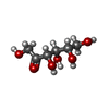  Type: L-saccharide / Mass: 180.156 Da / Num. of mol.: 5 Type: L-saccharide / Mass: 180.156 Da / Num. of mol.: 5Source method: isolated from a genetically manipulated source Formula: C6H12O6 #4: Sugar | ChemComp-SOE /   Type: L-saccharide, alpha linking / Mass: 180.156 Da / Num. of mol.: 4 Type: L-saccharide, alpha linking / Mass: 180.156 Da / Num. of mol.: 4Source method: isolated from a genetically manipulated source Formula: C6H12O6 #5: Water | ChemComp-HOH / |  Mass: 18.015 Da / Num. of mol.: 368 / Source method: isolated from a natural source / Formula: H2O Mass: 18.015 Da / Num. of mol.: 368 / Source method: isolated from a natural source / Formula: H2O |
|---|
-Experimental details
-Experiment
| Experiment | Method: X-RAY DIFFRACTION / Number of used crystals: 1 |
|---|
- Sample preparation
| Crystal | Density Matthews: 2.09 Å3/Da / Density % sol: 41.06 % |
|---|---|
| Crystal grow | Temperature: 293 K / Method: vapor diffusion, sitting drop / pH: 4.5 Details: Crystals of the His116Leu mutant of SR containing 10 mM NADPH and 100 mM L-sorbose were obtained under a reservoir solution condition of 30% (w/v) PEG400, 200 mM calcium chloride and 100 mM ...Details: Crystals of the His116Leu mutant of SR containing 10 mM NADPH and 100 mM L-sorbose were obtained under a reservoir solution condition of 30% (w/v) PEG400, 200 mM calcium chloride and 100 mM sodium acetate trihydrate, pH 4.5. The crystals of SR complexed with NADPH and L-sorbose were prepared by soaking the crystals in the reservoir solution supplemented with 2 M L-sorbose and 10 mM NADPH for 12 h at 293 K., VAPOR DIFFUSION, SITTING DROP, temperature 293.0K |
-Data collection
| Diffraction source | Source: SYNCHROTRON / Site: Photon Factory  / Beamline: AR-NE3A / Wavelength: 1 Å / Beamline: AR-NE3A / Wavelength: 1 Å |
|---|---|
| Detector | Type: ADSC QUANTUM 270 / Detector: CCD / Date: Oct 30, 2009 |
| Radiation | Protocol: SINGLE WAVELENGTH / Monochromatic (M) / Laue (L): M / Scattering type: x-ray |
| Radiation wavelength | Wavelength: 1 Å / Relative weight: 1 |
| Reflection | Resolution: 1.8→20 Å / Num. obs: 88419 / % possible obs: 99.8 % / Observed criterion σ(F): 1.0001 / Observed criterion σ(I): 1 / Rmerge(I) obs: 0.083 / Net I/σ(I): 21.3 |
| Reflection shell | Resolution: 1.8→1.85 Å / Rmerge(I) obs: 0.465 / Mean I/σ(I) obs: 5.6 / % possible all: 100 |
- Processing
| Software |
| |||||||||||||||||||||||||||||||||||||||||||||||||||||||||||||||||
|---|---|---|---|---|---|---|---|---|---|---|---|---|---|---|---|---|---|---|---|---|---|---|---|---|---|---|---|---|---|---|---|---|---|---|---|---|---|---|---|---|---|---|---|---|---|---|---|---|---|---|---|---|---|---|---|---|---|---|---|---|---|---|---|---|---|---|
| Refinement | Method to determine structure: MOLECULAR REPLACEMENT Starting model: PDB ENTRY 3AI2 Resolution: 1.8→19.69 Å / Cor.coef. Fo:Fc: 0.924 / Cor.coef. Fo:Fc free: 0.885 / SU B: 2.367 / SU ML: 0.076 / Cross valid method: THROUGHOUT / ESU R: 0.162 / ESU R Free: 0.146 / Stereochemistry target values: MAXIMUM LIKELIHOOD / Details: HYDROGENS HAVE BEEN ADDED IN THE RIDING POSITIONS
| |||||||||||||||||||||||||||||||||||||||||||||||||||||||||||||||||
| Solvent computation | Solvent model: NONE PARAMETERS FOR MASK CACLULATION | |||||||||||||||||||||||||||||||||||||||||||||||||||||||||||||||||
| Displacement parameters | Biso mean: 12.871 Å2
| |||||||||||||||||||||||||||||||||||||||||||||||||||||||||||||||||
| Refinement step | Cycle: LAST / Resolution: 1.8→19.69 Å
| |||||||||||||||||||||||||||||||||||||||||||||||||||||||||||||||||
| Refine LS restraints |
| |||||||||||||||||||||||||||||||||||||||||||||||||||||||||||||||||
| LS refinement shell | Resolution: 1.8→1.847 Å / Total num. of bins used: 20
|
