 Movie
Movie Controller

[English] 日本語
 Yorodumi
- PDB-3abo: Crystal structure of ethanolamine ammonia-lyase from Escherichia ... -
+ Open data

- Basic information
| Entry | Database: PDB / ID: 3abo | |||||||||
|---|---|---|---|---|---|---|---|---|---|---|
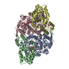
| Title | Crystal structure of ethanolamine ammonia-lyase from Escherichia coli complexed with CN-Cbl and ethanolamine | |||||||||
 Components Components | (Ethanolamine ammonia-lyase ...) x 2 | |||||||||
 Keywords Keywords | LYASE / (beta/alpha)8 fold / Cobalt / Cobalamin | |||||||||
| Function / homology |  Function and homology information Function and homology informationethanolamine ammonia-lyase / ethanolamine ammonia-lyase activity / ethanolamine ammonia-lyase complex / ethanolamine degradation polyhedral organelle / ethanolamine catabolic process / cobalamin binding / amino acid metabolic process / cytosol Similarity search - Function | |||||||||
| Biological species |  | |||||||||
| Method |  X-RAY DIFFRACTION / SYNCHROTRON / SAD, molecular replacement / Resolution: 2.1 Å X-RAY DIFFRACTION / SYNCHROTRON / SAD, molecular replacement / Resolution: 2.1 Å | |||||||||
 Authors Authors | Shibata, N. | |||||||||
 Citation Citation | Journal: J.Biol.Chem. / Year: 2010 Title: Crystal structures of ethanolamine ammonia-lyase complexed with coenzyme B12 analogs and substrates. Authors: Shibata, N. / Tamagaki, H. / Hieda, N. / Akita, K. / Komori, H. / Shomura, Y. / Terawaki, S. / Mori, K. / Yasuoka, N. / Higuchi, Y. / Toraya, T. | |||||||||
| History |
|
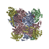
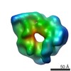
- Structure visualization
| Structure viewer | Molecule: MolmilJmol/JSmol |
|---|
- Downloads & links
-Download
| PDBx/mmCIF format | 3abo.cif.gz | 566.3 KB | Display | PDBx/mmCIF format |
|---|---|---|---|---|
| PDB format | pdb3abo.ent.gz | 466.8 KB | Display | PDB format |
| PDBx/mmJSON format | 3abo.json.gz | Tree view | PDBx/mmJSON format | |
| Others |  Other downloads Other downloads |
-Validation report
| Arichive directory | https://data.pdbj.org/pub/pdb/validation_reports/ab/3aboftp://data.pdbj.org/pub/pdb/validation_reports/ab/3abo | HTTPS FTP |
|---|
-Related structure data
| Related structure data |  3abqC  3abrC  3absC 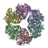 2qezS C: citing same article ( S: Starting model for refinement |
|---|---|
| Similar structure data |
-Links
 PDBj
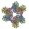
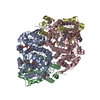
PDBj- Assembly
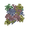
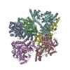
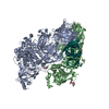
| Deposited unit | 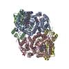
| ||||||||||||||||||
|---|---|---|---|---|---|---|---|---|---|---|---|---|---|---|---|---|---|---|---|
| 1 | 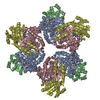
| ||||||||||||||||||
| 2 |
| ||||||||||||||||||
| Unit cell |
| ||||||||||||||||||
| Noncrystallographic symmetry (NCS) | NCS domain:
|
-Components
-Ethanolamine ammonia-lyase ... , 2 types, 4 molecules ACBD
| #1: Protein | Mass: 49447.816 Da / Num. of mol.: 2 Source method: isolated from a genetically manipulated source Source: (gene. exp.) #2: Protein | Mass: 33197.031 Da / Num. of mol.: 2 Source method: isolated from a genetically manipulated source Source: (gene. exp.) |
|---|
-Non-polymers , 5 types, 718 molecules 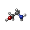


| #3: Chemical |  Type: L-peptide COOH carboxy terminus / Mass: 61.083 Da / Num. of mol.: 2 / Source method: obtained synthetically / Formula: C2H7NO Type: L-peptide COOH carboxy terminus / Mass: 61.083 Da / Num. of mol.: 2 / Source method: obtained synthetically / Formula: C2H7NO#4: Chemical | ChemComp-GOL /  Mass: 92.094 Da / Num. of mol.: 5 / Source method: obtained synthetically / Formula: C3H8O3 Mass: 92.094 Da / Num. of mol.: 5 / Source method: obtained synthetically / Formula: C3H8O3#5: Chemical |  Mass: 1330.356 Da / Num. of mol.: 2 / Source method: obtained synthetically / Formula: C62H89CoN13O14P Mass: 1330.356 Da / Num. of mol.: 2 / Source method: obtained synthetically / Formula: C62H89CoN13O14P#6: Chemical |  Mass: 22.990 Da / Num. of mol.: 2 / Source method: obtained synthetically / Formula: Na Mass: 22.990 Da / Num. of mol.: 2 / Source method: obtained synthetically / Formula: Na#7: Water | ChemComp-HOH / | Mass: 18.015 Da / Num. of mol.: 707 / Source method: isolated from a natural source / Formula: H2O |
|---|
-Experimental details
-Experiment
| Experiment | Method: X-RAY DIFFRACTION / Number of used crystals: 1 |
|---|
- Sample preparation
| Crystal | Density Matthews: 3.93 Å3/Da / Density % sol: 68.74 % |
|---|---|
| Crystal grow | Temperature: 277 K / Method: vapor diffusion, sitting drop / pH: 7 Details: 6.0-7.0 % (w/v) PEG 6000, 24-26 % (v/v) glycerol, 5.0 % (v/v) 2-propanol, 0.1 M HEPES-NaOH, pH 7.0, VAPOR DIFFUSION, SITTING DROP, temperature 277K |
-Data collection
| Diffraction | Mean temperature: 100 K |
|---|---|
| Diffraction source | Source: SYNCHROTRON / Site: SPring-8  / Beamline: BL38B1 / Wavelength: 1 Å / Beamline: BL38B1 / Wavelength: 1 Å |
| Detector | Type: RIGAKU JUPITER 210 / Detector: CCD / Date: Jul 11, 2008 / Details: mirrors |
| Radiation | Protocol: SINGLE WAVELENGTH / Monochromatic (M) / Laue (L): M / Scattering type: x-ray |
| Radiation wavelength | Wavelength: 1 Å / Relative weight: 1 |
| Reflection | Resolution: 2.1→50 Å / Num. all: 145836 / Num. obs: 145836 / % possible obs: 97.2 % / Observed criterion σ(I): 0 / Redundancy: 6.8 % / Rmerge(I) obs: 0.096 / Net I/σ(I): 17.9 |
| Reflection shell | Resolution: 2.1→2.15 Å / Redundancy: 6.3 % / Mean I/σ(I) obs: 4.35 / Num. unique all: 9228 / % possible all: 93.1 |
- Processing
| Software |
| ||||||||||||||||||||||||||||||||||||||||||||||||||||||||||||||||||||||||||||||||||||||||||||||||||||||||||||||||||||||||||||||||||||||||||||||||||||||||||||||||||||||||||
|---|---|---|---|---|---|---|---|---|---|---|---|---|---|---|---|---|---|---|---|---|---|---|---|---|---|---|---|---|---|---|---|---|---|---|---|---|---|---|---|---|---|---|---|---|---|---|---|---|---|---|---|---|---|---|---|---|---|---|---|---|---|---|---|---|---|---|---|---|---|---|---|---|---|---|---|---|---|---|---|---|---|---|---|---|---|---|---|---|---|---|---|---|---|---|---|---|---|---|---|---|---|---|---|---|---|---|---|---|---|---|---|---|---|---|---|---|---|---|---|---|---|---|---|---|---|---|---|---|---|---|---|---|---|---|---|---|---|---|---|---|---|---|---|---|---|---|---|---|---|---|---|---|---|---|---|---|---|---|---|---|---|---|---|---|---|---|---|---|---|---|---|
| Refinement | Method to determine structure: SAD, molecular replacement Starting model: PDB ENTRY 2QEZ Resolution: 2.1→47.51 Å / Cor.coef. Fo:Fc: 0.908 / Cor.coef. Fo:Fc free: 0.896 / SU B: 10.138 / SU ML: 0.127 / Cross valid method: THROUGHOUT / ESU R: 0.184 / ESU R Free: 0.167 / Stereochemistry target values: MAXIMUM LIKELIHOOD
| ||||||||||||||||||||||||||||||||||||||||||||||||||||||||||||||||||||||||||||||||||||||||||||||||||||||||||||||||||||||||||||||||||||||||||||||||||||||||||||||||||||||||||
| Solvent computation | Ion probe radii: 0.8 Å / Shrinkage radii: 0.8 Å / VDW probe radii: 1.2 Å / Solvent model: BABINET MODEL WITH MASK | ||||||||||||||||||||||||||||||||||||||||||||||||||||||||||||||||||||||||||||||||||||||||||||||||||||||||||||||||||||||||||||||||||||||||||||||||||||||||||||||||||||||||||
| Displacement parameters | Biso mean: 15.448 Å2
| ||||||||||||||||||||||||||||||||||||||||||||||||||||||||||||||||||||||||||||||||||||||||||||||||||||||||||||||||||||||||||||||||||||||||||||||||||||||||||||||||||||||||||
| Refinement step | Cycle: LAST / Resolution: 2.1→47.51 Å
| ||||||||||||||||||||||||||||||||||||||||||||||||||||||||||||||||||||||||||||||||||||||||||||||||||||||||||||||||||||||||||||||||||||||||||||||||||||||||||||||||||||||||||
| Refine LS restraints |
| ||||||||||||||||||||||||||||||||||||||||||||||||||||||||||||||||||||||||||||||||||||||||||||||||||||||||||||||||||||||||||||||||||||||||||||||||||||||||||||||||||||||||||
| Refine LS restraints NCS | Refine-ID: X-RAY DIFFRACTION
| ||||||||||||||||||||||||||||||||||||||||||||||||||||||||||||||||||||||||||||||||||||||||||||||||||||||||||||||||||||||||||||||||||||||||||||||||||||||||||||||||||||||||||
| LS refinement shell | Resolution: 2.1→2.155 Å / Total num. of bins used: 20
| ||||||||||||||||||||||||||||||||||||||||||||||||||||||||||||||||||||||||||||||||||||||||||||||||||||||||||||||||||||||||||||||||||||||||||||||||||||||||||||||||||||||||||
| Refinement TLS params. | Method: refined / Refine-ID: X-RAY DIFFRACTION
| ||||||||||||||||||||||||||||||||||||||||||||||||||||||||||||||||||||||||||||||||||||||||||||||||||||||||||||||||||||||||||||||||||||||||||||||||||||||||||||||||||||||||||
| Refinement TLS group |
|
