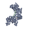
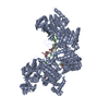
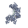
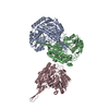
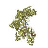
B: DNA (5'-D(*DTP*DTP*DTP*DTP*DTP*DCP*DCP*DCP*DAP*DCP*DCP*DTP*DTP*DTP*DT)-3') C: DNA (5'-D(P*DGP*DGP*DTP*DGP*DGP*DG)-3') E: DNA (5'-D(*DTP*DTP*DTP*DTP*DTP*DCP*DCP*DCP*DAP*DCP*DCP*DTP*DTP*DTP*DT)-3') F: DNA (5'-D(P*DGP*DGP*DTP*DGP*DGP*DG)-3') A: Protein recA D: Protein recA ヘテロ分子
pH: 8 詳細: the RecA5 fusion protein was incubated with a 1.5-fold molar excess of the primary d(T5C3AC2T4) oligonucleotide in protein buffer supplemented with 2 mM ADP, 10 mM MgCl2 and 8 mM AlF4, pH 6. ...詳細: the RecA5 fusion protein was incubated with a 1.5-fold molar excess of the primary d(T5C3AC2T4) oligonucleotide in protein buffer supplemented with 2 mM ADP, 10 mM MgCl2 and 8 mM AlF4, pH 6.0, for 30 min, followed by the addition of 2-fold molar excess of the complementary d(G2TG3) oligonucleotide. The crystals grew from 50 mM Tris-Cl, 9% (w/v) PVP K15, 32% (v/v) MPD, 100 mM magnesium acetate, 10 mM DTT, pH 8.0.
溶液の組成
ID
名称
Crystal-ID
Sol-ID
1
ADP
1
1
2
MgCl2
1
1
3
AlF4
1
1
4
Tris-Cl
1
1
5
PVPK15
1
1
6
MPD
1
1
7
magnesiumacetate
1
1
8
10mMDTT
1
1
9
Tris-Cl
1
2
10
PVPK15
1
2
11
MPD
1
2
12
magnesiumacetate
1
2
13
10mMDTT
1
2
-
データ収集
放射光源
由来: シンクロトロン / サイト: APS / ビームライン: 24-ID-C
検出器
日付: 2007年7月1日
放射
プロトコル: SINGLE WAVELENGTH / 単色(M)・ラウエ(L): M / 散乱光タイプ: x-ray
放射波長
相対比: 1
反射
解像度: 3.15→40 Å / Num. obs: 59390
-
解析
ソフトウェア
名称: REFMAC / バージョン: 5.3.0036 / 分類: 精密化
精密化
構造決定の手法: 分子置換 / 解像度: 3.15→40 Å / Cor.coef. Fo:Fc: 0.92 / Cor.coef. Fo:Fc free: 0.902 / SU B: 45.573 / SU ML: 0.373 / 交差検証法: THROUGHOUT / ESU R Free: 0.521 / 立体化学のターゲット値: MAXIMUM LIKELIHOOD
Rfactor
反射数
%反射
Selection details
Rfree
0.243
1231
2 %
RANDOM
Rwork
0.217
-
-
-
obs
0.218
59390
89.9 %
-
溶媒の処理
イオンプローブ半径: 0.8 Å / 減衰半径: 0.8 Å / VDWプローブ半径: 1.2 Å / 溶媒モデル: BABINET MODEL WITH MASK
 ムービー
ムービー コントローラー


 データを開く
 基本情報
基本情報 要素
要素 キーワード
キーワード 機能・相同性情報
 X線回折 /
  データ登録者
 引用
引用 構造の表示
構造の表示 ダウンロードとリンク
 その他のダウンロード


 PDBj
PDBj


 集合体
集合体


 分子量: 24.305 Da / 分子数: 10 / 由来タイプ: 合成 / 式: Mg
 分子量: 102.975 Da / 分子数: 10 / 由来タイプ: 合成 / 式: AlF4
 分子量: 427.201 Da / 分子数: 10 / 由来タイプ: 合成 / 式: C10H15N5O10P2 / コメント: ADP, エネルギー貯蔵分子*YM
 試料調製
試料調製 / ビームライン: 24-ID-C
 解析
解析