Movie
 Controller


+ Open data

- Basic information
| Entry | Database: PDB / ID: 38ht | |||||||||||||||
|---|---|---|---|---|---|---|---|---|---|---|---|---|---|---|---|---|
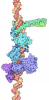
| Title | Cryo-EM structure of apo-pSTAT1 | |||||||||||||||
 Components | Signal transducer and activator of transcription 1-alpha/beta | |||||||||||||||
 Keywords | TRANSCRIPTION / Signal transducer and activator of transcription / transcription factor / Cytoplasm and Nuclear localization | |||||||||||||||
| Function / homology |  Function and homology information renal tubule development / metanephric mesenchymal cell differentiation / negative regulation of metanephric nephron tubule epithelial cell differentiation / negative regulation by virus of viral protein levels in host cell / negative regulation of mesenchymal to epithelial transition involved in metanephros morphogenesis / ISGF3 complex / response to interferon-beta / metanephric mesenchymal cell proliferation involved in metanephros development / interleukin-27-mediated signaling pathway / Differentiation of naive CD4+ T cells to T helper 1 cells (Th1 cells) ...renal tubule development / metanephric mesenchymal cell differentiation / negative regulation of metanephric nephron tubule epithelial cell differentiation / negative regulation by virus of viral protein levels in host cell / negative regulation of mesenchymal to epithelial transition involved in metanephros morphogenesis / ISGF3 complex / response to interferon-beta / metanephric mesenchymal cell proliferation involved in metanephros development / interleukin-27-mediated signaling pathway / Differentiation of naive CD4+ T cells to T helper 1 cells (Th1 cells) / Interleukin-9 signaling / Interleukin-21 signaling / interleukin-7-mediated signaling pathway / interleukin-9-mediated signaling pathway / positive regulation of mesenchymal cell proliferation / tumor necrosis factor receptor binding / Interleukin-27 signaling / Interleukin-35 Signalling / Signaling by cytosolic FGFR1 fusion mutants / cell surface receptor signaling pathway via STAT / NOTCH3 Intracellular Domain Regulates Transcription / Interleukin-20 family signaling / Interleukin-6 signaling / type I interferon-mediated signaling pathway / negative regulation of endothelial cell proliferation / histone acetyltransferase binding / Regulation of IFNA/IFNB signaling / positive regulation of interferon-alpha production / ubiquitin-like protein ligase binding / Regulation of IFNG signaling / type II interferon-mediated signaling pathway / Growth hormone receptor signaling / RNA polymerase II core promoter sequence-specific DNA binding / cellular response to interferon-beta / cell surface receptor signaling pathway via JAK-STAT / positive regulation of defense response to virus by host / Signaling by PDGFRA transmembrane, juxtamembrane and kinase domain mutants / Signaling by PDGFRA extracellular domain mutants / Signaling by CSF3 (G-CSF) / negative regulation of angiogenesis / tumor necrosis factor-mediated signaling pathway / response to type II interferon / positive regulation of erythrocyte differentiation / Downstream signal transduction / Turbulent (oscillatory, disturbed) flow shear stress activates signaling by PIEZO1 and integrins in endothelial cells / defense response / transcription corepressor binding / Signaling by phosphorylated juxtamembrane, extracellular and kinase domain KIT mutants / negative regulation of canonical NF-kappaB signal transduction / RNA polymerase II transcription regulatory region sequence-specific DNA binding / promoter-specific chromatin binding / cellular response to type II interferon / Downregulation of SMAD2/3:SMAD4 transcriptional activity / Signaling by SCF-KIT / Inactivation of CSF3 (G-CSF) signaling / PKR-mediated signaling / response to peptide hormone / ISG15 antiviral mechanism / Interferon gamma signaling / RNA polymerase II transcription regulator complex / transcription coactivator binding / Signaling by CSF1 (M-CSF) in myeloid cells / Interferon alpha/beta signaling / Regulation of RUNX2 expression and activity / Signaling by ALK fusions and activated point mutants / histone binding / Regulation of PD-L1(CD274) transcription / defense response to virus / regulation of cell population proliferation / Interleukin-4 and Interleukin-13 signaling / double-stranded DNA binding / regulation of apoptotic process / DNA-binding transcription factor activity, RNA polymerase II-specific / RNA polymerase II cis-regulatory region sequence-specific DNA binding / cadherin binding / DNA-binding transcription factor activity / regulation of transcription by RNA polymerase II / positive regulation of DNA-templated transcription / chromatin / negative regulation of transcription by RNA polymerase II / SARS-CoV-2 activates/modulates innate and adaptive immune responses / perinuclear region of cytoplasm / enzyme binding / positive regulation of transcription by RNA polymerase II / protein homodimerization activity / protein-containing complex / nucleoplasm / identical protein binding / nucleus / cytosol / cytoplasm Similarity search - Function | |||||||||||||||
| Biological species |  Homo sapiens (human) | |||||||||||||||
| Method | ELECTRON MICROSCOPY / single particle reconstruction / cryo EM / Resolution: 3.3 Å | |||||||||||||||
 Authors | Kumar, P. / Bellis, N.F. / Vinkemeier, U. / Cingolani, G. | |||||||||||||||
| Funding support |  United States, 4items
| |||||||||||||||
 Citation | Journal: bioRxiv / Year: 2026 Title: Cytokine-induced nuclear translocation of STAT1 via a non-transferable NLS. Authors: Pramod Kumar / James R Cavey / Ruoyu Yang / Oran White / Andreas Begitt / Liu He / Stephanie S Suinn / Nathan F Bellis / Ravi K Lokareddy / Uwe Vinkemeier / Gino Cingolani /  Abstract: The targeting function of nuclear localization signals (NLSs) is generally considered independent of a protein's native sequence or fold and is readily transferable to heterologous cargos. Contrary ...The targeting function of nuclear localization signals (NLSs) is generally considered independent of a protein's native sequence or fold and is readily transferable to heterologous cargos. Contrary to this paradigm, rapid nuclear translocation of phosphorylated STAT1 (pSTAT1) following cytokine stimulation requires importin β, Ran-GTP, and the importin α5 isoform, which recognizes a non-transferable NLS. Here, we present cryo-EM structures of pSTAT1 bound to importin α5, revealing an asymmetric 2:1 complex that diverges from canonical NLS-mediated cargo recognition. Importin α5 occupies the DNA-binding groove of the pSTAT1 dimer, with a single STAT1 N-terminal domain positioning the C-terminal Armadillo repeats 9-10 (S1B domain) orthogonal to the DNA-binding interface. This interface is also targeted by the Ebola virus protein VP24, an antagonist of interferon signaling. We further show that Ran-GTP alone is insufficient to trigger nuclear release of pSTAT1, which additionally requires the exportin CAS. A cryo-EM reconstruction of the CAS-Ran-GTP-α5 complex, supported by in vitro competition assays, demonstrates that CAS and pSTAT1 are mutually exclusive ligands for importin α5. Together, these findings define the molecular choreography of cytokine-induced STAT1 nuclear translocation and release, establishing a general paradigm for STAT family signaling. | |||||||||||||||
| History |
|
- Structure visualization
| Structure viewer | Molecule: MolmilJmol/JSmol |
|---|
- Downloads & links
-Download
| PDBx/mmCIF format | 38ht.cif.gz | 397.1 KB | Display | PDBx/mmCIF format |
|---|---|---|---|---|
| PDB format | pdb38ht.ent.gz | 324.7 KB | Display | PDB format |
| PDBx/mmJSON format | 38ht.json.gz | Tree view | PDBx/mmJSON format | |
| Others |  Other downloads |
-Validation report
| Arichive directory | https://data.pdbj.org/pub/pdb/validation_reports/8h/38htftp://data.pdbj.org/pub/pdb/validation_reports/8h/38ht | HTTPS FTP |
|---|
-Related structure data
| Related structure data |  78826MC  11jgC  11phC  38ykC  75683 11gr C: citing same article ( M: map data used to model this data |
|---|---|
| Similar structure data |


-Links
 PDBj
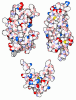
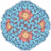
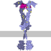
- Assembly
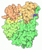
| Deposited unit | 
|
|---|---|
| 1 |
|
-Components
| #1: Protein | Mass: 77696.773 Da / Num. of mol.: 2 Source method: isolated from a genetically manipulated source Details: STAT1 / Source: (gene. exp.) Homo sapiens (human) / Gene: STAT1 / Plasmid: pAcPNV / Production host:   Spodoptera frugiperda (fall armyworm) / References: UniProt: P42224 Has ligand of interest | Y | Has protein modification | Y | |
|---|
-Experimental details
-Experiment
| Experiment | Method: ELECTRON MICROSCOPY |
|---|---|
| EM experiment | Aggregation state: PARTICLE / 3D reconstruction method: single particle reconstruction |
- Sample preparation
| Component | Name: This is a dimeric assembly of two phosphorylated STAT1 core protomers. Type: COMPLEX / Entity ID: all / Source: RECOMBINANT |
|---|---|
| Molecular weight | Value: 0.071750 MDa / Experimental value: NO |
| Source (natural) | Organism: Homo sapiens (human) |
| Source (recombinant) | Organism: Spodoptera frugiperda (fall armyworm) / Plasmid: pAcPNV |
| Buffer solution | pH: 8 Details: 20mM Tris, pH 8.0, 150 mM NaCl, 3mM Beta Mercapto Ethanol, 1mM phenylmethylsulfonyl fluoride. |
| Specimen | Conc.: 1.5 mg/ml / Embedding applied: NO / Shadowing applied: NO / Staining applied: NO / Vitrification applied: YES / Details: The sample was mono disperse. |
| Specimen support | Grid material: GOLD / Grid mesh size: 300 divisions/in. / Grid type: UltrAuFoil R1.2/1.3 |
| Vitrification | Instrument: FEI VITROBOT MARK IV / Cryogen name: ETHANE / Humidity: 100 % / Chamber temperature: 295 K |
- Electron microscopy imaging
| Experimental equipment | 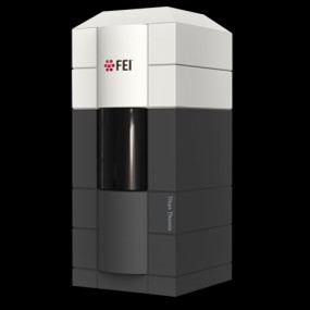 Model: Titan Krios / Image courtesy: FEI Company |
|---|---|
| Microscopy | Model: TFS KRIOS |
| Electron gun | Electron source:  FIELD EMISSION GUN / Accelerating voltage: 300 kV / Illumination mode: SPOT SCAN |
| Electron lens | Mode: BRIGHT FIELD / Nominal magnification: 105000 X / Nominal defocus max: 2500 nm / Nominal defocus min: 800 nm / Cs: 2.7 mm / C2 aperture diameter: 70 µm / Alignment procedure: COMA FREE |
| Specimen holder | Cryogen: NITROGEN / Specimen holder model: FEI TITAN KRIOS AUTOGRID HOLDER |
| Image recording | Average exposure time: 2.98 sec. / Electron dose: 50 e/Å2 / Film or detector model: GATAN K3 BIOQUANTUM (6k x 4k) / Num. of grids imaged: 1 / Num. of real images: 19493 |
- Processing
| EM software |
| ||||||||||||||||||||||||||||||||||||
|---|---|---|---|---|---|---|---|---|---|---|---|---|---|---|---|---|---|---|---|---|---|---|---|---|---|---|---|---|---|---|---|---|---|---|---|---|---|
| CTF correction | Type: PHASE FLIPPING AND AMPLITUDE CORRECTION | ||||||||||||||||||||||||||||||||||||
| Particle selection | Num. of particles selected: 6483490 | ||||||||||||||||||||||||||||||||||||
| Symmetry | Point symmetry: C2 (2 fold cyclic) | ||||||||||||||||||||||||||||||||||||
| 3D reconstruction | Resolution: 3.3 Å / Resolution method: FSC 0.143 CUT-OFF / Num. of particles: 196697 / Symmetry type: POINT | ||||||||||||||||||||||||||||||||||||
| Atomic model building | B value: 125 / Protocol: RIGID BODY FIT / Space: REAL / Target criteria: Cross correlation | ||||||||||||||||||||||||||||||||||||
| Atomic model building | PDB-ID: 11GR 11gr Accession code: 11GR / Chain residue range: 133-716 / Pdb chain residue range: 133-716 / Source name: PDB / Type: experimental model |