 Movie
Movie Controller

[English] 日本語
 Yorodumi
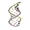
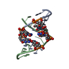
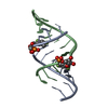
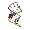
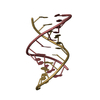
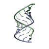
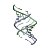
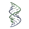
- PDB-353d: CRYSTAL STRUCTURE OF DOMAIN A OF THERMUS FLAVUS 5S RRNA AND THE C... -
+ Open data

- Basic information
| Entry | Database: PDB / ID: 353d | ||||||
|---|---|---|---|---|---|---|---|
| Title | CRYSTAL STRUCTURE OF DOMAIN A OF THERMUS FLAVUS 5S RRNA AND THE CONTRIBUTION OF WATER MOLECULES TO ITS STRUCTURE | ||||||
 Components Components |
| ||||||
 Keywords Keywords | RNA / A-RNA / DOUBLE STRAND | ||||||
| Function / homology | RNA / RNA (> 10) Function and homology information Function and homology information | ||||||
| Method |  X-RAY DIFFRACTION / SYNCHROTRON / Resolution: 2.4 Å X-RAY DIFFRACTION / SYNCHROTRON / Resolution: 2.4 Å | ||||||
 Authors Authors | Betzel, C. / Lorenz, S. / Furste, J.P. / Bald, R. / Zhang, M. / Schneider, T.R. / Wilson, K.S. / Erdmann, V.A. | ||||||
 Citation Citation | Journal: FEBS Lett. / Year: 1994 Title: Crystal structure of domain A of Thermus flavus 5S rRNA and the contribution of water molecules to its structure. Authors: Betzel, C. / Lorenz, S. / Furste, J.P. / Bald, R. / Zhang, M. / Schneider, T.R. / Wilson, K.S. / Erdmann, V.A. #1: Journal: To be PublishedTitle: Structure of Domain A of Thermus flavus 5S rRNA at 2.3 A Resolution. Comparison of Nearest Neighbor Parameters for Mismatches and the Influence of Solvent Authors: Perbandt, M. / Lorenz, S. / Betzel, C. / Erdmann, V.A. #2: Journal: Acta Crystallogr.,Sect.D / Year: 1993Title: Crystallization and Preliminary Diffraction Studies of the Chemically Synthesized Domain A of Thermus flavus 5S rRNA: An Dodecamer Double Helix Authors: Lorenz, S. / Furste, J. / Bald, R. / Zhang, M. / Raderschall, E. / Betzel, C. / Dauter, Z. / Wilson, K. | ||||||
| History |
|
- Structure visualization
| Structure viewer | Molecule: MolmilJmol/JSmol |
|---|
- Downloads & links
-Download
| PDBx/mmCIF format | 353d.cif.gz | 26.7 KB | Display | PDBx/mmCIF format |
|---|---|---|---|---|
| PDB format | pdb353d.ent.gz | 17.9 KB | Display | PDB format |
| PDBx/mmJSON format | 353d.json.gz | Tree view | PDBx/mmJSON format | |
| Others |  Other downloads Other downloads |
-Validation report
| Arichive directory | https://data.pdbj.org/pub/pdb/validation_reports/53/353dftp://data.pdbj.org/pub/pdb/validation_reports/53/353d | HTTPS FTP |
|---|
-Related structure data
| Related structure data |  1rnaS S: Starting model for refinement |
|---|---|
| Similar structure data |


-Links
 PDBj
PDBj


- Assembly
| Deposited unit | 
| ||||||||
|---|---|---|---|---|---|---|---|---|---|
| 1 |
| ||||||||
| Unit cell |
|
-Components
| #1: RNA chain | Mass: 3723.264 Da / Num. of mol.: 1 / Source method: obtained synthetically |
|---|---|
| #2: RNA chain | Mass: 3963.408 Da / Num. of mol.: 1 / Source method: obtained synthetically |
| #3: Water | ChemComp-HOH /  Mass: 18.015 Da / Num. of mol.: 158 / Source method: isolated from a natural source / Formula: H2O Mass: 18.015 Da / Num. of mol.: 158 / Source method: isolated from a natural source / Formula: H2O |
-Experimental details
-Experiment
| Experiment | Method: X-RAY DIFFRACTION / Number of used crystals: 2 |
|---|
- Sample preparation
| Crystal | Density Matthews: 2.6 Å3/Da / Density % sol: 51.91 % | ||||||||||||||||||||||||||||||||||||||||||||||||||||||
|---|---|---|---|---|---|---|---|---|---|---|---|---|---|---|---|---|---|---|---|---|---|---|---|---|---|---|---|---|---|---|---|---|---|---|---|---|---|---|---|---|---|---|---|---|---|---|---|---|---|---|---|---|---|---|---|
| Crystal grow | Temperature: 291 K / Method: vapor diffusion, hanging drop / pH: 6.5 Details: pH 6.50, VAPOR DIFFUSION, HANGING DROP, temperature 291K | ||||||||||||||||||||||||||||||||||||||||||||||||||||||
| Components of the solutions |
| ||||||||||||||||||||||||||||||||||||||||||||||||||||||
| Crystal grow | *PLUS Temperature: 45 ℃ / pH: 6.5 Details: Lorenz, S., (1993) Acta Crystallogr.,Sect.D, 49, 418. | ||||||||||||||||||||||||||||||||||||||||||||||||||||||
| Components of the solutions | *PLUS
|
-Data collection
| Diffraction |
| |||||||||||||||||||||
|---|---|---|---|---|---|---|---|---|---|---|---|---|---|---|---|---|---|---|---|---|---|---|
| Diffraction source |
| |||||||||||||||||||||
| Detector |
| |||||||||||||||||||||
| Radiation |
| |||||||||||||||||||||
| Radiation wavelength |
| |||||||||||||||||||||
| Reflection | Highest resolution: 2.4 Å / Num. obs: 2477 / % possible obs: 83.5 % | |||||||||||||||||||||
| Reflection | *PLUS Highest resolution: 2.4 Å / % possible obs: 83.5 % |

- Processing
| Software |
| |||||||||||||||||||||||||||||||||||||||||||||||||||||||||||||||
|---|---|---|---|---|---|---|---|---|---|---|---|---|---|---|---|---|---|---|---|---|---|---|---|---|---|---|---|---|---|---|---|---|---|---|---|---|---|---|---|---|---|---|---|---|---|---|---|---|---|---|---|---|---|---|---|---|---|---|---|---|---|---|---|---|
| Refinement | Starting model: NDB ENTRY ARN035 (PDB ENTRY 1RNA) Resolution: 2.4→8 Å / σ(F): 0 / Details: X-PLOR (BRUNGER) ALSO WAS USED. /
| |||||||||||||||||||||||||||||||||||||||||||||||||||||||||||||||
| Displacement parameters | Biso mean: 23.9 Å2 | |||||||||||||||||||||||||||||||||||||||||||||||||||||||||||||||
| Refine Biso |
| |||||||||||||||||||||||||||||||||||||||||||||||||||||||||||||||
| Refinement step | Cycle: LAST / Resolution: 2.4→8 Å
| |||||||||||||||||||||||||||||||||||||||||||||||||||||||||||||||
| Refine LS restraints |
| |||||||||||||||||||||||||||||||||||||||||||||||||||||||||||||||
| LS refinement shell | Resolution: 2.3→2.4 Å / % reflection obs: 50 % | |||||||||||||||||||||||||||||||||||||||||||||||||||||||||||||||
| Software | *PLUS Name: NUCLSQ / Classification: refinement | |||||||||||||||||||||||||||||||||||||||||||||||||||||||||||||||
| Refinement | *PLUS Highest resolution: 2.4 Å / Lowest resolution: 8 Å / σ(F): 0 / Rfactor obs: 0.18 / Rfactor Rwork: 0.18 | |||||||||||||||||||||||||||||||||||||||||||||||||||||||||||||||
| Solvent computation | *PLUS | |||||||||||||||||||||||||||||||||||||||||||||||||||||||||||||||
| Displacement parameters | *PLUS Biso mean: 23.9 Å2 | |||||||||||||||||||||||||||||||||||||||||||||||||||||||||||||||
| Refine LS restraints | *PLUS
|
