[English] 日本語
Yorodumi
- PDB-32cr: SHANK3 PDZ (570-664) in complex with an internal PDZ binding moti... -

+
Open data


ID or keywords:

Loading...

-
Basic information

Entry
Database: PDB / ID: 32cr
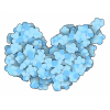
TitleSHANK3 PDZ (570-664) in complex with an internal PDZ binding motif (843-864) in Densin-180
Components
  • Leucine-rich repeat-containing protein 7
  • SH3 and multiple ankyrin repeat domains protein 3
KeywordsPROTEIN BINDING / Postsynaptic density PDZ domain
Function / homology
Function and homology information


response to toluene / response to interleukin-17 / regulation of AMPA glutamate receptor clustering / Neurexins and neuroligins / guanylate kinase-associated protein clustering / striatal medium spiny neuron differentiation / positive regulation of synapse structural plasticity / synaptic receptor adaptor activity / sensory perception of touch / postsynaptic density assembly ...response to toluene / response to interleukin-17 / regulation of AMPA glutamate receptor clustering / Neurexins and neuroligins / guanylate kinase-associated protein clustering / striatal medium spiny neuron differentiation / positive regulation of synapse structural plasticity / synaptic receptor adaptor activity / sensory perception of touch / postsynaptic density assembly / embryonic epithelial tube formation / vocal learning / regulation of grooming behavior / structural constituent of postsynaptic density / negative regulation of actin filament bundle assembly / neurotransmitter receptor transport, endosome to postsynaptic membrane / NMDA glutamate receptor clustering / positive regulation of long-term neuronal synaptic plasticity / RET signaling / negative regulation of cell volume / establishment or maintenance of epithelial cell apical/basal polarity / brain morphogenesis / dendritic spine morphogenesis / neuron spine / regulation of dendritic spine morphogenesis / vocalization behavior / axon initial segment / regulation of behavioral fear response / regulation of long-term synaptic potentiation / neural precursor cell proliferation / neuromuscular process controlling balance / long-term synaptic depression / locomotion / exploration behavior / RAF/MAP kinase cascade / regulation of postsynapse organization / Neutrophil degranulation / ciliary membrane / adult behavior / positive regulation of dendritic spine development / positive regulation of glutamate receptor signaling pathway / receptor clustering / AMPA glutamate receptor clustering / associative learning / locomotory exploration behavior / social behavior / Unblocking of NMDA receptors, glutamate binding and activation / glial cell proliferation / postsynaptic density, intracellular component / regulation of long-term synaptic depression / positive regulation of synaptic transmission, glutamatergic / synapse assembly / ionotropic glutamate receptor binding / learning / excitatory synapse / positive regulation of excitatory postsynaptic potential / positive regulation of long-term synaptic potentiation / locomotory behavior / filopodium / adherens junction / positive regulation of neuron projection development / cell-cell adhesion / regulation of synaptic plasticity / memory / SH3 domain binding / synapse organization / postsynaptic density membrane / G protein-coupled receptor binding / modulation of chemical synaptic transmission / long-term synaptic potentiation / gene expression / MAPK cascade / actin cytoskeleton / actin binding / scaffold protein binding / dendritic spine / basolateral plasma membrane / learning or memory / neuron projection / postsynaptic density / protein kinase binding / protein-containing complex binding / glutamatergic synapse / zinc ion binding / identical protein binding / plasma membrane / cytosol / cytoplasm
Similarity search - Function
: / PDZ domain 6 / : / PDZ domain / : / Leucine-rich repeat region / Variant SH3 domain / SAM domain (Sterile alpha motif) / Leucine-rich repeats, bacterial type / Leucine-rich repeat, SDS22-like subfamily ...: / PDZ domain 6 / : / PDZ domain / : / Leucine-rich repeat region / Variant SH3 domain / SAM domain (Sterile alpha motif) / Leucine-rich repeats, bacterial type / Leucine-rich repeat, SDS22-like subfamily / SAM domain profile. / Sterile alpha motif. / Sterile alpha motif domain / Leucine rich repeat / Leucine-rich repeat, typical subtype / Leucine-rich repeats, typical (most populated) subfamily / Sterile alpha motif/pointed domain superfamily / Leucine-rich repeat profile. / PDZ domain / PDZ domain profile. / Domain present in PSD-95, Dlg, and ZO-1/2. / PDZ domain / Leucine-rich repeat / PDZ superfamily / Ankyrin repeat profile. / Ankyrin repeats (3 copies) / Ankyrin repeat region circular profile. / ankyrin repeats / Leucine-rich repeat domain superfamily / Ankyrin repeat / Src homology 3 domains / Ankyrin repeat-containing domain superfamily / SH3-like domain superfamily / Src homology 3 (SH3) domain profile. / SH3 domain
Similarity search - Domain/homology
Leucine-rich repeat-containing protein 7 / SH3 and multiple ankyrin repeat domains protein 3
Similarity search - Component
Biological speciesMus musculus (house mouse)
Rattus norvegicus (Norway rat)
MethodX-RAY DIFFRACTION / SYNCHROTRON / MOLECULAR REPLACEMENT / Resolution: 1.98 Å
AuthorsKallem, T. / Goult, B.T.
Funding support United Kingdom, 1items
OrganizationGrant numberCountry
British Heart FoundationSP/F/23/150045 United Kingdom
CitationJournal: Biorxiv / Year: 2026
Title: An internal PDZ-binding motif in Densin-180 promotes activity-dependent SHANK scaffold remodelling
Authors: Otani, Y. / Srinivasan, V. / Toller, J. / Kallem, T. / Ball, N. / Barsukov, I. / Saarikangas, J. / Kreienkamp, H.J. / Goult, B.T.
History
DepositionJul 5, 2026Deposition site: PDBE / Processing site: PDBE
Revision 1.0Jul 22, 2026Provider: repository / Type: Initial release

-
Structure visualization

Structure viewerMolecule:
MolmilJmol/JSmol

Downloads & links

-
Assembly

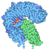
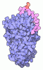
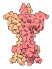
Deposited unit
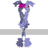
A: SH3 and multiple ankyrin repeat domains protein 3
B: Leucine-rich repeat-containing protein 7
C: SH3 and multiple ankyrin repeat domains protein 3
D: Leucine-rich repeat-containing protein 7
E: SH3 and multiple ankyrin repeat domains protein 3
F: Leucine-rich repeat-containing protein 7
G: SH3 and multiple ankyrin repeat domains protein 3
H: Leucine-rich repeat-containing protein 7


Theoretical massNumber of molelcules
Total (without water)54,7468
Polymers54,7468
Non-polymers00
Water6,612367
1
A: SH3 and multiple ankyrin repeat domains protein 3
B: Leucine-rich repeat-containing protein 7
G: SH3 and multiple ankyrin repeat domains protein 3
H: Leucine-rich repeat-containing protein 7


Theoretical massNumber of molelcules
Total (without water)27,3734
Polymers27,3734
Non-polymers00
Water724
TypeNameSymmetry operationNumber
identity operation1_555x,y,z1
Buried area3920 Å2
ΔGint-17 kcal/mol
Surface area12220 Å2
MethodPISA
2
C: SH3 and multiple ankyrin repeat domains protein 3
D: Leucine-rich repeat-containing protein 7

E: SH3 and multiple ankyrin repeat domains protein 3
F: Leucine-rich repeat-containing protein 7


Theoretical massNumber of molelcules
Total (without water)27,3734
Polymers27,3734
Non-polymers00
Water724
TypeNameSymmetry operationNumber
identity operation1_555x,y,z1
crystal symmetry operation2_554-x+1/2,-y,z-1/21
Buried area3810 Å2
ΔGint-20 kcal/mol
Surface area12170 Å2
MethodPISA
Unit cell
Length a, b, c (Å)63.89, 71.34, 114.92
Angle α, β, γ (deg.)90, 90, 90
Int Tables number19
Space group name H-MP212121

-
Components

#1: Protein
SH3 and multiple ankyrin repeat domains protein 3 / Shank3 / Proline-rich synapse-associated protein 2 / ProSAP2 / SPANK-2


Mass: 10971.632 Da / Num. of mol.: 4
Source method: isolated from a genetically manipulated source
Source: (gene. exp.) Mus musculus (house mouse) / Gene: Shank3, Kiaa1650, Prosap2 / Production host: Escherichia coli (E. coli) / References: UniProt: Q4ACU6
#2: Protein/peptide
Leucine-rich repeat-containing protein 7 / Densin-180 / Densin / Protein LAP1


Mass: 2714.980 Da / Num. of mol.: 4 / Source method: obtained synthetically / Source: (synth.) Rattus norvegicus (Norway rat) / References: UniProt: P70587
#3: Water ChemComp-HOH / water


Mass: 18.015 Da / Num. of mol.: 367 / Source method: isolated from a natural source / Formula: H2O
Has protein modificationY

-
Experimental details

-
Experiment

ExperimentMethod: X-RAY DIFFRACTION / Number of used crystals: 1

-
Sample preparation

CrystalDensity Matthews: 2.39 Å3/Da / Density % sol: 48.58 %
Crystal growTemperature: 292.15 K / Method: vapor diffusion, sitting drop / pH: 8.5
Details: 60 mM magnesium chloride hexahydrate, 60 mM calcium chloride dihydrate, 100 mM Tris/Bicine pH 8.5, 12% v/v PEG 500 MME, 6% w/v PEG 20000

-
Data collection

DiffractionMean temperature: 100 K / Serial crystal experiment: N
Diffraction sourceSource: SYNCHROTRON / Site: Diamond / Beamline: I24 / Wavelength: 0.62 Å
DetectorType: DECTRIS EIGER2 X 9M / Detector: PIXEL / Date: Jun 25, 2026
RadiationProtocol: SINGLE WAVELENGTH / Monochromatic (M) / Laue (L): M / Scattering type: x-ray
Radiation wavelengthWavelength: 0.62 Å / Relative weight: 1
ReflectionResolution: 1.98→47.64 Å / Num. obs: 37309 / % possible obs: 100 % / Redundancy: 6.6 % / CC1/2: 1 / Rrim(I) all: 0.201 / Net I/σ(I): 7.6
Reflection shellResolution: 1.98→2.01 Å / Redundancy: 5.7 % / Mean I/σ(I) obs: 1.1 / Num. unique obs: 1820 / CC1/2: 0.4 / Rrim(I) all: 1.804 / % possible all: 99.8

-
Processing

Software
NameVersionClassification
REFMAC5.8.0430 (refmacat 0.4.105)refinement
xia23diidata reduction
xia23diidata scaling
MOLREPphasing
RefinementMethod to determine structure: MOLECULAR REPLACEMENT / Resolution: 1.98→47.639 Å / Cor.coef. Fo:Fc: 0.955 / Cor.coef. Fo:Fc free: 0.926 / SU B: 6.239 / SU ML: 0.161 / Cross valid method: FREE R-VALUE / ESU R: 0.18 / ESU R Free: 0.172
Details: Hydrogens have been added in their riding positions
RfactorNum. reflection% reflection
Rfree0.2566 1727 4.635 %
Rwork0.201 35533 -
all0.204 --
obs-37260 99.89 %
Solvent computationIon probe radii: 0.8 Å / Shrinkage radii: 0.8 Å / VDW probe radii: 1.2 Å / Solvent model: MASK BULK SOLVENT
Displacement parametersBiso mean: 39.889 Å2
Baniso -1Baniso -2Baniso -3
1--1.55 Å2-0 Å20 Å2
2--1.401 Å20 Å2
3---0.149 Å2
Refinement stepCycle: LAST / Resolution: 1.98→47.639 Å
ProteinNucleic acidLigandSolventTotal
Num. atoms3524 0 0 367 3891
Refine LS restraints
Refine-IDTypeDev idealDev ideal targetNumber
X-RAY DIFFRACTIONr_bond_refined_d0.0080.0123640
X-RAY DIFFRACTIONr_bond_other_d0.0010.0163568
X-RAY DIFFRACTIONr_angle_refined_deg1.7281.834951
X-RAY DIFFRACTIONr_angle_other_deg0.5811.758186
X-RAY DIFFRACTIONr_dihedral_angle_1_deg6.6445468
X-RAY DIFFRACTIONr_dihedral_angle_2_deg8.428528
X-RAY DIFFRACTIONr_dihedral_angle_3_deg15.62210592
X-RAY DIFFRACTIONr_dihedral_angle_6_deg15.49910155
X-RAY DIFFRACTIONr_chiral_restr0.0830.2571
X-RAY DIFFRACTIONr_gen_planes_refined0.0080.024305
X-RAY DIFFRACTIONr_gen_planes_other0.0010.02847
X-RAY DIFFRACTIONr_nbd_refined0.2060.2645
X-RAY DIFFRACTIONr_symmetry_nbd_other0.2010.23280
X-RAY DIFFRACTIONr_nbtor_refined0.1810.21707
X-RAY DIFFRACTIONr_symmetry_nbtor_other0.0880.21934
X-RAY DIFFRACTIONr_xyhbond_nbd_refined0.20.2295
X-RAY DIFFRACTIONr_symmetry_xyhbond_nbd_other0.0780.25
X-RAY DIFFRACTIONr_symmetry_nbd_refined0.2170.221
X-RAY DIFFRACTIONr_nbd_other0.2080.260
X-RAY DIFFRACTIONr_symmetry_xyhbond_nbd_refined0.1530.210
X-RAY DIFFRACTIONr_mcbond_it4.0113.8241839
X-RAY DIFFRACTIONr_mcbond_other4.013.8241840
X-RAY DIFFRACTIONr_mcangle_it5.9136.8092285
X-RAY DIFFRACTIONr_mcangle_other5.9126.812286
X-RAY DIFFRACTIONr_scbond_it4.3224.251801
X-RAY DIFFRACTIONr_scbond_other4.324.251802
X-RAY DIFFRACTIONr_scangle_it6.5437.7142655
X-RAY DIFFRACTIONr_scangle_other6.5427.7142656
X-RAY DIFFRACTIONr_lrange_it9.29139.4523952
X-RAY DIFFRACTIONr_lrange_other9.27238.5483870
LS refinement shell
Resolution (Å)Rfactor RfreeNum. reflection RfreeRfactor RworkNum. reflection RworkRefine-ID% reflection obs (%)
1.98-2.0310.3671170.3372580X-RAY DIFFRACTION99.778
2.031-2.0870.3331160.32522X-RAY DIFFRACTION99.7731
2.087-2.1470.3361080.2922461X-RAY DIFFRACTION99.8057
2.147-2.2130.3161240.2722387X-RAY DIFFRACTION99.8807
2.213-2.2860.277910.2322334X-RAY DIFFRACTION99.9588
2.286-2.3660.2931200.2342229X-RAY DIFFRACTION99.9575
2.366-2.4550.3061030.222191X-RAY DIFFRACTION99.9129
2.455-2.5550.261110.2122065X-RAY DIFFRACTION99.9541
2.555-2.6680.2651050.192004X-RAY DIFFRACTION99.9526
2.668-2.7980.2471030.1911894X-RAY DIFFRACTION99.95
2.798-2.9490.227910.1791833X-RAY DIFFRACTION100
2.949-3.1270.245850.1891739X-RAY DIFFRACTION99.9452
3.127-3.3420.267890.1981623X-RAY DIFFRACTION100
3.342-3.6080.243710.1891546X-RAY DIFFRACTION99.9382
3.608-3.950.225780.1711409X-RAY DIFFRACTION100
3.95-4.4120.206570.1591298X-RAY DIFFRACTION99.9263
4.412-5.0880.214590.1431154X-RAY DIFFRACTION100
5.088-6.2130.194360.172997X-RAY DIFFRACTION99.8068
6.213-8.7130.247400.195787X-RAY DIFFRACTION99.5187
8.713-47.6390.384230.225480X-RAY DIFFRACTION99.604

+
About Yorodumi

-
News

-
Feb 9, 2022. New format data for meta-information of EMDB entries

New format data for meta-information of EMDB entries

  • Version 3 of the EMDB header file is now the official format.
  • The previous official version 1.9 will be removed from the archive.

Related info.:EMDB header

External links:wwPDB to switch to version 3 of the EMDB data model

-
Aug 12, 2020. Covid-19 info

Covid-19 info

URL: https://pdbj.org/emnavi/covid19.php

New page: Covid-19 featured information page in EM Navigator.

Related info.:Covid-19 info / Mar 5, 2020. Novel coronavirus structure data

+
Mar 5, 2020. Novel coronavirus structure data

Novel coronavirus structure data

Related info.:Yorodumi Speices / Aug 12, 2020. Covid-19 info

External links:COVID-19 featured content - PDBj / Molecule of the Month (242):Coronavirus Proteases

+
Jan 31, 2019. EMDB accession codes are about to change! (news from PDBe EMDB page)

EMDB accession codes are about to change! (news from PDBe EMDB page)

  • The allocation of 4 digits for EMDB accession codes will soon come to an end. Whilst these codes will remain in use, new EMDB accession codes will include an additional digit and will expand incrementally as the available range of codes is exhausted. The current 4-digit format prefixed with “EMD-” (i.e. EMD-XXXX) will advance to a 5-digit format (i.e. EMD-XXXXX), and so on. It is currently estimated that the 4-digit codes will be depleted around Spring 2019, at which point the 5-digit format will come into force.
  • The EM Navigator/Yorodumi systems omit the EMD- prefix.

Related info.:Q: What is EMD? / ID/Accession-code notation in Yorodumi/EM Navigator

External links:EMDB Accession Codes are Changing Soon! / Contact to PDBj

+
Jul 12, 2017. Major update of PDB

Major update of PDB

  • wwPDB released updated PDB data conforming to the new PDBx/mmCIF dictionary.
  • This is a major update changing the version number from 4 to 5, and with Remediation, in which all the entries are updated.
  • In this update, many items about electron microscopy experimental information are reorganized (e.g. em_software).
  • Now, EM Navigator and Yorodumi are based on the updated data.

External links:wwPDB Remediation / Enriched Model Files Conforming to OneDep Data Standards Now Available in the PDB FTP Archive

-
Yorodumi

Thousand views of thousand structures

  • Yorodumi is a browser for structure data from EMDB, PDB, SASBDB, etc.
  • This page is also the successor to EM Navigator detail page, and also detail information page/front-end page for Omokage search.
  • The word "yorodu" (or yorozu) is an old Japanese word meaning "ten thousand". "mi" (miru) is to see.

Related info.:EMDB / PDB / SASBDB / Comparison of 3 databanks / Yorodumi Search / Aug 31, 2016. New EM Navigator & Yorodumi / Yorodumi Papers / Jmol/JSmol / Function and homology information / Changes in new EM Navigator and Yorodumi

Read more