 Movie
Movie Controller

[English] 日本語
 Yorodumi
- PDB-2zyt: Crystal structure of mouse cytosolic sulfotransferase mSULT1D1 co... -
+ Open data

- Basic information
| Entry | Database: PDB / ID: 2zyt | ||||||
|---|---|---|---|---|---|---|---|
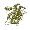
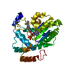
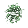
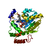
| Title | Crystal structure of mouse cytosolic sulfotransferase mSULT1D1 complex with PAPS | ||||||
 Components Components | Tyrosine-ester sulfotransferase | ||||||
 Keywords Keywords | TRANSFERASE / SULT1D1 / SULT / sulfotransferase / PAPS / sulfonation | ||||||
| Function / homology |  Function and homology information Function and homology informationTransferases; Transferring sulfur-containing groups; Sulfotransferases / aryl sulfotransferase activity / sulfation / sulfotransferase activity / sulfate assimilation / catecholamine metabolic process / lipid metabolic process / cytoplasm Similarity search - Function | ||||||
| Biological species |  | ||||||
| Method |  X-RAY DIFFRACTION / SYNCHROTRON / MOLECULAR REPLACEMENT / Resolution: 1.55 Å X-RAY DIFFRACTION / SYNCHROTRON / MOLECULAR REPLACEMENT / Resolution: 1.55 Å | ||||||
 Authors Authors | Teramoto, T. / Sakakibara, Y. / Liu, M.-C. / Suiko, M. / Kimura, M. / Kakuta, Y. | ||||||
 Citation Citation | Journal: Biochem.Biophys.Res.Commun. / Year: 2009 Title: Snapshot of a Michaelis complex in a sulfuryl transfer reaction: Crystal structure of a mouse sulfotransferase, mSULT1D1, complexed with donor substrate and accepter substrate Authors: Teramoto, T. / Sakakibara, Y. / Liu, M.-C. / Suiko, M. / Kimura, M. / Kakuta, Y. | ||||||
| History |
|
- Structure visualization
| Structure viewer | Molecule: MolmilJmol/JSmol |
|---|
- Downloads & links
-Download
| PDBx/mmCIF format | 2zyt.cif.gz | 82.4 KB | Display | PDBx/mmCIF format |
|---|---|---|---|---|
| PDB format | pdb2zyt.ent.gz | 60 KB | Display | PDB format |
| PDBx/mmJSON format | 2zyt.json.gz | Tree view | PDBx/mmJSON format | |
| Others |  Other downloads Other downloads |
-Validation report
| Arichive directory | https://data.pdbj.org/pub/pdb/validation_reports/zy/2zytftp://data.pdbj.org/pub/pdb/validation_reports/zy/2zyt | HTTPS FTP |
|---|
-Related structure data
| Related structure data |  2zyuC  2zyvC  2zywC  2zptS S: Starting model for refinement C: citing same article ( |
|---|---|
| Similar structure data |


-Links
 PDBj
PDBj
- Assembly
| Deposited unit | 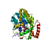
| ||||||||
|---|---|---|---|---|---|---|---|---|---|
| 1 |
| ||||||||
| Unit cell |
|
-Components
| #1: Protein | Mass: 35128.293 Da / Num. of mol.: 1 / Fragment: residues 1-295 Source method: isolated from a genetically manipulated source Source: (gene. exp.)  References: UniProt: Q9R2C2, UniProt: Q3UZZ6*PLUS, EC: 2.8.2.9 | ||
|---|---|---|---|
| #2: Chemical | ChemComp-PPS /   Mass: 507.264 Da / Num. of mol.: 1 / Source method: obtained synthetically / Formula: C10H15N5O13P2S Mass: 507.264 Da / Num. of mol.: 1 / Source method: obtained synthetically / Formula: C10H15N5O13P2S | ||
| #3: Chemical |   Mass: 92.094 Da / Num. of mol.: 2 / Source method: obtained synthetically / Formula: C3H8O3 Mass: 92.094 Da / Num. of mol.: 2 / Source method: obtained synthetically / Formula: C3H8O3#4: Water | ChemComp-HOH / |  Mass: 18.015 Da / Num. of mol.: 229 / Source method: isolated from a natural source / Formula: H2O Mass: 18.015 Da / Num. of mol.: 229 / Source method: isolated from a natural source / Formula: H2O |
-Experimental details
-Experiment
| Experiment | Method: X-RAY DIFFRACTION / Number of used crystals: 1 |
|---|
- Sample preparation
| Crystal | Density Matthews: 3.37 Å3/Da / Density % sol: 63.51 % |
|---|---|
| Crystal grow | Temperature: 293 K / Method: vapor diffusion, hanging drop / pH: 5.5 Details: 16% PEG 10000, 10mM dithiothreitol, 100mM Bis-Tris, pH 5.5, VAPOR DIFFUSION, HANGING DROP, temperature 293K |
-Data collection
| Diffraction | Mean temperature: 100 K |
|---|---|
| Diffraction source | Source: SYNCHROTRON / Site: SPring-8  / Beamline: BL38B1 / Wavelength: 1 Å / Beamline: BL38B1 / Wavelength: 1 Å |
| Detector | Type: RIGAKU JUPITER 210 / Detector: CCD / Details: mirrors |
| Radiation | Monochromator: Si 111 monochromater / Protocol: SINGLE WAVELENGTH / Monochromatic (M) / Laue (L): M / Scattering type: x-ray |
| Radiation wavelength | Wavelength: 1 Å / Relative weight: 1 |
| Reflection | Resolution: 1.55→50 Å / Num. all: 55335 / Num. obs: 55335 / % possible obs: 81.4 % / Redundancy: 3.2 % / Rmerge(I) obs: 0.093 / Rsym value: 0.093 / Net I/σ(I): 14.2 |
| Reflection shell | Resolution: 1.55→1.61 Å / Redundancy: 2.5 % / Rmerge(I) obs: 0.527 / Mean I/σ(I) obs: 1.2 / Num. unique all: 2890 / Rsym value: 0.527 / % possible all: 42.8 |
- Processing
| Software |
| ||||||||||||||||||||||||||||||||||||||||||||||||||||||||||||||||||||||||||||||||||||||||||
|---|---|---|---|---|---|---|---|---|---|---|---|---|---|---|---|---|---|---|---|---|---|---|---|---|---|---|---|---|---|---|---|---|---|---|---|---|---|---|---|---|---|---|---|---|---|---|---|---|---|---|---|---|---|---|---|---|---|---|---|---|---|---|---|---|---|---|---|---|---|---|---|---|---|---|---|---|---|---|---|---|---|---|---|---|---|---|---|---|---|---|---|
| Refinement | Method to determine structure: MOLECULAR REPLACEMENT Starting model: PDB ENTRY 2ZPT Resolution: 1.55→34.67 Å / Cor.coef. Fo:Fc: 0.962 / Cor.coef. Fo:Fc free: 0.955 / SU B: 1.39 / SU ML: 0.051 / Cross valid method: THROUGHOUT / ESU R: 0.084 / ESU R Free: 0.082 / Stereochemistry target values: MAXIMUM LIKELIHOOD / Details: HYDROGENS HAVE BEEN ADDED IN THE RIDING POSITIONS
| ||||||||||||||||||||||||||||||||||||||||||||||||||||||||||||||||||||||||||||||||||||||||||
| Solvent computation | Ion probe radii: 0.8 Å / Shrinkage radii: 0.8 Å / VDW probe radii: 1.2 Å / Solvent model: MASK | ||||||||||||||||||||||||||||||||||||||||||||||||||||||||||||||||||||||||||||||||||||||||||
| Displacement parameters | Biso mean: 22.943 Å2
| ||||||||||||||||||||||||||||||||||||||||||||||||||||||||||||||||||||||||||||||||||||||||||
| Refinement step | Cycle: LAST / Resolution: 1.55→34.67 Å
| ||||||||||||||||||||||||||||||||||||||||||||||||||||||||||||||||||||||||||||||||||||||||||
| Refine LS restraints |
| ||||||||||||||||||||||||||||||||||||||||||||||||||||||||||||||||||||||||||||||||||||||||||
| LS refinement shell | Resolution: 1.548→1.588 Å / Total num. of bins used: 20
|
