 Movie
Movie Controller

[English] 日本語
 Yorodumi
- PDB-2zxf: Crystal structure of human glycyl-trna synthetase (GLYRS) in comp... -
+ Open data

- Basic information
| Entry | Database: PDB / ID: 2zxf | ||||||
|---|---|---|---|---|---|---|---|
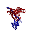
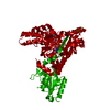
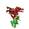
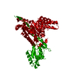
| Title | Crystal structure of human glycyl-trna synthetase (GLYRS) in complex with AP4A (cocrystallized with AP4A) | ||||||
 Components Components | Glycyl-tRNA synthetase | ||||||
 Keywords Keywords | LIGASE / AP4A / glycine / ATP / GLY-AMP / tRNA / aminoacyl-tRNA synthetase / ATP-binding / charcot-marie-tooth disease / disease mutation / nucleotide-binding / phosphoprotein / protein biosynthesis | ||||||
| Function / homology |  Function and homology information Function and homology informationmitochondrial glycyl-tRNA aminoacylation / ATP:ATP adenylyltransferase activity / glycine-tRNA ligase / glycine-tRNA ligase activity / bis(5'-nucleosyl)-tetraphosphatase (asymmetrical) activity / Mitochondrial tRNA aminoacylation / diadenosine tetraphosphate biosynthetic process / Cytosolic tRNA aminoacylation / tRNA aminoacylation for protein translation / secretory granule ...mitochondrial glycyl-tRNA aminoacylation / ATP:ATP adenylyltransferase activity / glycine-tRNA ligase / glycine-tRNA ligase activity / bis(5'-nucleosyl)-tetraphosphatase (asymmetrical) activity / Mitochondrial tRNA aminoacylation / diadenosine tetraphosphate biosynthetic process / Cytosolic tRNA aminoacylation / tRNA aminoacylation for protein translation / secretory granule / Transferases; Transferring phosphorus-containing groups; Nucleotidyltransferases / protein dimerization activity / mitochondrial matrix / axon / mitochondrion / extracellular exosome / ATP binding / identical protein binding / cytoplasm / cytosol Similarity search - Function | ||||||
| Biological species |  Homo sapiens (human) Homo sapiens (human) | ||||||
| Method |  X-RAY DIFFRACTION / SYNCHROTRON / MOLECULAR REPLACEMENT / Resolution: 3.4 Å X-RAY DIFFRACTION / SYNCHROTRON / MOLECULAR REPLACEMENT / Resolution: 3.4 Å | ||||||
 Authors Authors | Guo, R.T. / Yang, X.L. / Schimmel, P. | ||||||
 Citation Citation | Journal: To be Published Title: Crystal structures and biochemical analyses suggest unique mechanism and role for human GlyRS in Ap4A homeostasis Authors: Guo, R.T. / Chong, Y.E. / Guo, M. / Yang, X.L. | ||||||
| History |
|
- Structure visualization
| Structure viewer | Molecule: MolmilJmol/JSmol |
|---|
- Downloads & links
-Download
| PDBx/mmCIF format | 2zxf.cif.gz | 125.1 KB | Display | PDBx/mmCIF format |
|---|---|---|---|---|
| PDB format | pdb2zxf.ent.gz | 95 KB | Display | PDB format |
| PDBx/mmJSON format | 2zxf.json.gz | Tree view | PDBx/mmJSON format | |
| Others |  Other downloads Other downloads |
-Validation report
| Arichive directory | https://data.pdbj.org/pub/pdb/validation_reports/zx/2zxfftp://data.pdbj.org/pub/pdb/validation_reports/zx/2zxf | HTTPS FTP |
|---|
-Related structure data
| Related structure data |  2zt5C  2zt6C  2zt7C  2zt8C  2pmeS S: Starting model for refinement C: citing same article ( |
|---|---|
| Similar structure data |


-Links
 PDBj
PDBj
- Assembly
| Deposited unit | 
| ||||||||
|---|---|---|---|---|---|---|---|---|---|
| 1 | 
| ||||||||
| Unit cell |
|
-Components
| #1: Protein | Mass: 78704.828 Da / Num. of mol.: 1 / Fragment: UNP residues 55-739 Source method: isolated from a genetically manipulated source Source: (gene. exp.) Homo sapiens (human) / Gene: GARS / Plasmid: pET21A / Production host:  |
|---|---|
| #2: Chemical | ChemComp-B4P /   Mass: 836.387 Da / Num. of mol.: 1 / Source method: obtained synthetically / Formula: C20H28N10O19P4 Mass: 836.387 Da / Num. of mol.: 1 / Source method: obtained synthetically / Formula: C20H28N10O19P4 |
| #3: Water | ChemComp-HOH /  Mass: 18.015 Da / Num. of mol.: 71 / Source method: isolated from a natural source / Formula: H2O Mass: 18.015 Da / Num. of mol.: 71 / Source method: isolated from a natural source / Formula: H2O |
-Experimental details
-Experiment
| Experiment | Method: X-RAY DIFFRACTION / Number of used crystals: 1 |
|---|
- Sample preparation
| Crystal | Density Matthews: 3.5 Å3/Da / Density % sol: 64.9 % |
|---|---|
| Crystal grow | Temperature: 298 K / Method: vapor diffusion, hanging drop / pH: 7.5 Details: 25mM Tris-HCL, 150mM NaCl, 10mM MgCl2, 4M Sodium formate, PH7.5, VAPOR DIFFUSION, HANGING DROP, temperature 298.0K |
-Data collection
| Diffraction | Mean temperature: 100 K |
|---|---|
| Diffraction source | Source: SYNCHROTRON / Site: SSRL  / Beamline: BL11-1 / Wavelength: 1 Å / Beamline: BL11-1 / Wavelength: 1 Å |
| Detector | Type: ADSC QUANTUM 315 / Detector: CCD / Date: May 20, 2008 / Details: mirrors |
| Radiation | Protocol: SINGLE WAVELENGTH / Monochromatic (M) / Laue (L): M / Scattering type: x-ray |
| Radiation wavelength | Wavelength: 1 Å / Relative weight: 1 |
| Reflection | Resolution: 3.4→50 Å / Num. all: 15555 / Num. obs: 14819 / % possible obs: 95.1 % / Observed criterion σ(F): 0 / Observed criterion σ(I): 0 / Redundancy: 6.6 % / Rmerge(I) obs: 0.043 / Net I/σ(I): 37.8 |
| Reflection shell | Resolution: 3.4→3.52 Å / Redundancy: 4.3 % / Rmerge(I) obs: 0.272 / Mean I/σ(I) obs: 4 / Num. unique all: 1536 / % possible all: 71.5 |
- Processing
| Software |
| |||||||||||||||||||||||||
|---|---|---|---|---|---|---|---|---|---|---|---|---|---|---|---|---|---|---|---|---|---|---|---|---|---|---|
| Refinement | Method to determine structure: MOLECULAR REPLACEMENT Starting model: PDB ENTRY 2PME Resolution: 3.4→50 Å / Isotropic thermal model: Isotropic / Cross valid method: THROUGHOUT / σ(F): 0 / σ(I): 0 / Stereochemistry target values: Engh & Huber
| |||||||||||||||||||||||||
| Displacement parameters | Biso mean: 174.9 Å2 | |||||||||||||||||||||||||
| Refine analyze | Luzzati sigma a obs: 1.3 Å | |||||||||||||||||||||||||
| Refinement step | Cycle: LAST / Resolution: 3.4→50 Å
| |||||||||||||||||||||||||
| Refine LS restraints |
| |||||||||||||||||||||||||
| LS refinement shell | Resolution: 3.4→3.52 Å
|
