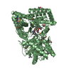
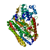
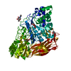
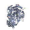
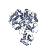
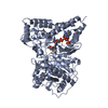
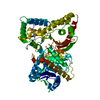
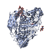
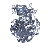
HYDROLASE / BETA-PRISM FOLD SURROUNDED BY SIX ALPHA HELIX / Lipid metabolism / Secreted
Function / homology
Function and homology information
sphingosine catabolic process / ceramidase / N-acylsphingosine amidohydrolase activity / ceramide catabolic process / sphingosine biosynthetic process / long-chain fatty acid biosynthetic process / extracellular region / membrane / metal ion binding Similarity search - Function
Mass: 18.015 Da / Num. of mol.: 939 / Source method: isolated from a natural source / Formula: H2O
-
Details
Has protein modification
Y
Sequence details
ABOUT THE CONFLICTS OF RESIDUE 157, 172 AND 574, REFERE TO UNP Q9I596 REFERENCE 1 AND THE GENBANK ...ABOUT THE CONFLICTS OF RESIDUE 157, 172 AND 574, REFERE TO UNP Q9I596 REFERENCE 1 AND THE GENBANK DATABASE BAA88409.
-
Experimental details
-
Experiment
Experiment
Method: X-RAY DIFFRACTION / Number of used crystals: 1
-
Sample preparation
Crystal
Density Matthews: 2.6 Å3/Da / Density % sol: 52.68 %
Crystal grow
Temperature: 293 K / Method: vapor diffusion, hanging drop / pH: 7.4 Details: 14% PEG3350, 200mM Ammonium Formate, 1mM C2-seramide, pH 7.4, VAPOR DIFFUSION, HANGING DROP, temperature 293K
In the structure databanks used in Yorodumi, some data are registered as the other names, "COVID-19 virus" and "2019-nCoV". Here are the details of the virus and the list of structure data.
Jan 31, 2019. EMDB accession codes are about to change! (news from PDBe EMDB page)
EMDB accession codes are about to change! (news from PDBe EMDB page)
The allocation of 4 digits for EMDB accession codes will soon come to an end. Whilst these codes will remain in use, new EMDB accession codes will include an additional digit and will expand incrementally as the available range of codes is exhausted. The current 4-digit format prefixed with “EMD-” (i.e. EMD-XXXX) will advance to a 5-digit format (i.e. EMD-XXXXX), and so on. It is currently estimated that the 4-digit codes will be depleted around Spring 2019, at which point the 5-digit format will come into force.
The EM Navigator/Yorodumi systems omit the EMD- prefix.
Related info.:Q: What is EMD? / ID/Accession-code notation in Yorodumi/EM Navigator
Yorodumi is a browser for structure data from EMDB, PDB, SASBDB, etc.
This page is also the successor to EM Navigator detail page, and also detail information page/front-end page for Omokage search.
The word "yorodu" (or yorozu) is an old Japanese word meaning "ten thousand". "mi" (miru) is to see.
Related info.:EMDB / PDB / SASBDB / Comparison of 3 databanks / Yorodumi Search / Aug 31, 2016. New EM Navigator & Yorodumi / Yorodumi Papers / Jmol/JSmol / Function and homology information / Changes in new EM Navigator and Yorodumi
 Movie
Movie Controller


 Open data
 Basic information
 Components
 Keywords
 Function and homology information
 Pseudomonas aeruginosa (bacteria)
 X-RAY DIFFRACTION /
  Authors
 Citation
 Structure visualization
 Downloads & links
 Other downloads
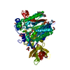

 PDBj
PDBj Assembly


 Mass: 65.409 Da / Num. of mol.: 2 / Source method: obtained synthetically / Formula: Zn
 Mass: 341.529 Da / Num. of mol.: 2 / Source method: obtained synthetically / Formula: C20H39NO3
 Mass: 46.025 Da / Num. of mol.: 8 / Source method: obtained synthetically / Formula: CH2O2
 Mass: 24.305 Da / Num. of mol.: 2 / Source method: obtained synthetically / Formula: Mg
 Mass: 78.133 Da / Num. of mol.: 2 / Source method: obtained synthetically / Formula: C2H6OS / Comment: DMSO, precipitant*YM
 Sample preparation
 / Beamline: BL41XU / Wavelength: 1 Å
 Processing