 Movie
Movie Controller

[English] 日本語
 Yorodumi
- PDB-2zu7: Crystal structure of mannosyl-3-phosphoglycerate synthase from Py... -
+ Open data

- Basic information
| Entry | Database: PDB / ID: 2zu7 | ||||||
|---|---|---|---|---|---|---|---|
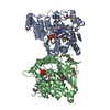
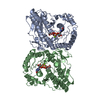
| Title | Crystal structure of mannosyl-3-phosphoglycerate synthase from Pyrococcus horikoshii | ||||||
 Components Components | Mannosyl-3-phosphoglycerate synthase | ||||||
 Keywords Keywords | TRANSFERASE / GT-A fold / glycosyltransferase / GT55 / Cytoplasm / Magnesium | ||||||
| Function / homology |  Function and homology information Function and homology informationmannosyl-3-phosphoglycerate synthase / mannosyl-3-phosphoglycerate synthase activity / mannosylglycerate biosynthetic process / cytoplasm Similarity search - Function | ||||||
| Biological species |   Pyrococcus horikoshii (archaea) Pyrococcus horikoshii (archaea) | ||||||
| Method |  X-RAY DIFFRACTION / SYNCHROTRON / SAD / Resolution: 2.5 Å X-RAY DIFFRACTION / SYNCHROTRON / SAD / Resolution: 2.5 Å | ||||||
 Authors Authors | Kawamura, T. / Watanabe, N. / Tanaka, I. | ||||||
 Citation Citation | Journal: To be Published Title: Crystal structure of mannosyl-3-phosphoglycerate synthase from Pyrococcus horikoshii Authors: Kawamura, T. / Watanabe, N. / Tanaka, I. | ||||||
| History |
|
- Structure visualization
| Structure viewer | Molecule: MolmilJmol/JSmol |
|---|
- Downloads & links
-Download
| PDBx/mmCIF format | 2zu7.cif.gz | 152.7 KB | Display | PDBx/mmCIF format |
|---|---|---|---|---|
| PDB format | pdb2zu7.ent.gz | 123.4 KB | Display | PDB format |
| PDBx/mmJSON format | 2zu7.json.gz | Tree view | PDBx/mmJSON format | |
| Others |  Other downloads Other downloads |
-Validation report
| Arichive directory | https://data.pdbj.org/pub/pdb/validation_reports/zu/2zu7ftp://data.pdbj.org/pub/pdb/validation_reports/zu/2zu7 | HTTPS FTP |
|---|
-Related structure data


-Links
 PDBj
PDBj

- Assembly
| Deposited unit | 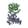
| ||||||||
|---|---|---|---|---|---|---|---|---|---|
| 1 |
| ||||||||
| Unit cell |
|
-Components
| #1: Protein | Mass: 45977.918 Da / Num. of mol.: 2 Source method: isolated from a genetically manipulated source Source: (gene. exp.) Pyrococcus horikoshii (archaea) / Strain: OT3 / Gene: PH0927 / Plasmid: pET22B / Production host:  References: UniProt: O58689, mannosyl-3-phosphoglycerate synthase #2: Water | ChemComp-HOH / |  Mass: 18.015 Da / Num. of mol.: 35 / Source method: isolated from a natural source / Formula: H2O Mass: 18.015 Da / Num. of mol.: 35 / Source method: isolated from a natural source / Formula: H2OHas protein modification | Y | |
|---|
-Experimental details
-Experiment
| Experiment | Method: X-RAY DIFFRACTION / Number of used crystals: 1 |
|---|
- Sample preparation
| Crystal | Density Matthews: 2.12 Å3/Da / Density % sol: 42.07 % |
|---|---|
| Crystal grow | Temperature: 293 K / Method: vapor diffusion, hanging drop / pH: 5.2 Details: 30% MPD, 0.05M sodium acetate, pH 5.2, VAPOR DIFFUSION, HANGING DROP, temperature 293K |
-Data collection
| Diffraction | Mean temperature: 95 K |
|---|---|
| Diffraction source | Source: SYNCHROTRON / Site: SPring-8  / Beamline: BL41XU / Wavelength: 0.9789 Å / Beamline: BL41XU / Wavelength: 0.9789 Å |
| Detector | Type: ADSC QUANTUM 315 / Detector: CCD / Date: Sep 29, 2007 |
| Radiation | Monochromator: double-crystal monochromator / Protocol: SINGLE WAVELENGTH / Monochromatic (M) / Laue (L): M / Scattering type: x-ray |
| Radiation wavelength | Wavelength: 0.9789 Å / Relative weight: 1 |
| Reflection | Resolution: 2.5→50 Å / Num. obs: 26230 / % possible obs: 97.6 % / Observed criterion σ(I): -3 / Redundancy: 4.1 % / Biso Wilson estimate: 38.8 Å2 / Rsym value: 0.07 / Net I/σ(I): 25.3 |
| Reflection shell | Resolution: 2.5→2.59 Å / Redundancy: 3.5 % / Mean I/σ(I) obs: 9.5 / Num. unique all: 2404 / Rsym value: 0.144 / % possible all: 91 |
- Processing
| Software |
| ||||||||||||||||||||||||||||||||||||||||||||||||||||||||||||||||||||||||||||||||
|---|---|---|---|---|---|---|---|---|---|---|---|---|---|---|---|---|---|---|---|---|---|---|---|---|---|---|---|---|---|---|---|---|---|---|---|---|---|---|---|---|---|---|---|---|---|---|---|---|---|---|---|---|---|---|---|---|---|---|---|---|---|---|---|---|---|---|---|---|---|---|---|---|---|---|---|---|---|---|---|---|---|
| Refinement | Method to determine structure: SAD / Resolution: 2.5→29.93 Å / Rfactor Rfree error: 0.007 / Data cutoff high absF: 1588512.95 / Data cutoff low absF: 0 / Isotropic thermal model: RESTRAINED / Cross valid method: THROUGHOUT / σ(F): 0 / Stereochemistry target values: Engh & Huber
| ||||||||||||||||||||||||||||||||||||||||||||||||||||||||||||||||||||||||||||||||
| Solvent computation | Solvent model: FLAT MODEL / Bsol: 23.9615 Å2 / ksol: 0.300717 e/Å3 | ||||||||||||||||||||||||||||||||||||||||||||||||||||||||||||||||||||||||||||||||
| Displacement parameters | Biso mean: 42.7 Å2
| ||||||||||||||||||||||||||||||||||||||||||||||||||||||||||||||||||||||||||||||||
| Refine analyze |
| ||||||||||||||||||||||||||||||||||||||||||||||||||||||||||||||||||||||||||||||||
| Refinement step | Cycle: LAST / Resolution: 2.5→29.93 Å
| ||||||||||||||||||||||||||||||||||||||||||||||||||||||||||||||||||||||||||||||||
| Refine LS restraints |
| ||||||||||||||||||||||||||||||||||||||||||||||||||||||||||||||||||||||||||||||||
| LS refinement shell | Resolution: 2.5→2.66 Å / Rfactor Rfree error: 0.022 / Total num. of bins used: 6
| ||||||||||||||||||||||||||||||||||||||||||||||||||||||||||||||||||||||||||||||||
| Xplor file |
|
