Redundancy: 6.8 % / Av σ(I) over netI: 26.46 / Number: 318115 / Rmerge(I) obs: 0.083 / Χ2: 1.68 / D res high: 2 Å / D res low: 50 Å / Num. obs: 46906 / % possible obs: 95.5
Diffraction reflection shell
Highest resolution (Å)
Lowest resolution (Å)
% possible obs (%)
ID
Rmerge(I) obs
Chi squared
Redundancy
2
2.07
73.4
1
0.509
0.815
4.2
2.07
2.15
86.7
1
0.385
0.855
5.3
2.15
2.25
95.5
1
0.326
0.901
6.2
2.25
2.37
99.7
1
0.283
0.926
7
2.37
2.52
100
1
0.225
1.029
7.4
2.52
2.71
100
1
0.166
1.212
7.5
2.71
2.99
100
1
0.12
1.469
7.5
2.99
3.42
100
1
0.078
1.968
7.5
3.42
4.31
100
1
0.054
2.69
7.4
4.31
50
99.8
1
0.05
3.84
7
Reflection
Resolution: 2→50 Å / Num. obs: 46906 / % possible obs: 95.5 % / Redundancy: 6.8 % / Rmerge(I) obs: 0.083 / Χ2: 1.679 / Net I/σ(I): 26.462
Reflection shell
Resolution (Å)
Redundancy (%)
Rmerge(I) obs
Num. unique all
Χ2
% possible all
2-2.07
4.2
0.509
3572
0.815
73.4
2.07-2.15
5.3
0.385
4231
0.855
86.7
2.15-2.25
6.2
0.326
4637
0.901
95.5
2.25-2.37
7
0.283
4856
0.926
99.7
2.37-2.52
7.4
0.225
4872
1.029
100
2.52-2.71
7.5
0.166
4911
1.212
100
2.71-2.99
7.5
0.12
4888
1.469
100
2.99-3.42
7.5
0.078
4936
1.968
100
3.42-4.31
7.4
0.054
4946
2.69
100
4.31-50
7
0.05
5057
3.84
99.8
-
Phasing
Phasing
Method: MAD
Phasing set
ID
1
2
Phasing MAD
D res high: 3 Å / D res low: 20 Å / FOM : 0.53 / Reflection: 14835
In the structure databanks used in Yorodumi, some data are registered as the other names, "COVID-19 virus" and "2019-nCoV". Here are the details of the virus and the list of structure data.
Jan 31, 2019. EMDB accession codes are about to change! (news from PDBe EMDB page)
EMDB accession codes are about to change! (news from PDBe EMDB page)
The allocation of 4 digits for EMDB accession codes will soon come to an end. Whilst these codes will remain in use, new EMDB accession codes will include an additional digit and will expand incrementally as the available range of codes is exhausted. The current 4-digit format prefixed with “EMD-” (i.e. EMD-XXXX) will advance to a 5-digit format (i.e. EMD-XXXXX), and so on. It is currently estimated that the 4-digit codes will be depleted around Spring 2019, at which point the 5-digit format will come into force.
The EM Navigator/Yorodumi systems omit the EMD- prefix.
Related info.:Q: What is EMD? / ID/Accession-code notation in Yorodumi/EM Navigator
Yorodumi is a browser for structure data from EMDB, PDB, SASBDB, etc.
This page is also the successor to EM Navigator detail page, and also detail information page/front-end page for Omokage search.
The word "yorodu" (or yorozu) is an old Japanese word meaning "ten thousand". "mi" (miru) is to see.
Related info.:EMDB / PDB / SASBDB / Comparison of 3 databanks / Yorodumi Search / Aug 31, 2016. New EM Navigator & Yorodumi / Yorodumi Papers / Jmol/JSmol / Function and homology information / Changes in new EM Navigator and Yorodumi
 Movie
Movie Controller

 Yorodumi
 Open data
 Basic information
 Components
 Keywords
 Function and homology information
 Pyrococcus horikoshii (archaea)
 X-RAY DIFFRACTION /
  Authors
 Citation
 Structure visualization
 Downloads & links
 Other downloads


 PDBj
PDBj
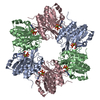
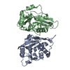
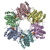
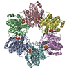
 Assembly


 Mass: 427.201 Da / Num. of mol.: 3 / Source method: obtained synthetically / Formula: C10H15N5O10P2 / Comment: ADP, energy-carrying molecule*YM
 Mass: 24.305 Da / Num. of mol.: 3 / Source method: obtained synthetically / Formula: Mg
 Mass: 18.015 Da / Num. of mol.: 88 / Source method: isolated from a natural source / Formula: H2O
 Sample preparation
 / Beamline: BL38B1 / Wavelength: 0.97905, 0.97934
 Processing