 Movie
Movie Controller

[English] 日本語
 Yorodumi
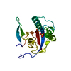
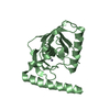
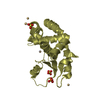
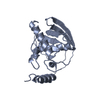
- PDB-2zr4: Crystal structure of a mutant PIN1 peptidyl-prolyl cis-trans isomerase -
+ Open data

- Basic information
| Entry | Database: PDB / ID: 2zr4 | ||||||
|---|---|---|---|---|---|---|---|
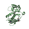
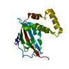
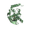
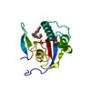
| Title | Crystal structure of a mutant PIN1 peptidyl-prolyl cis-trans isomerase | ||||||
 Components Components | Peptidyl-prolyl cis-trans isomerase NIMA-interacting 1 | ||||||
 Keywords Keywords | ISOMERASE / PIN1 mutant (S32A) / Cell cycle / Nucleus / Phosphoprotein / Rotamase | ||||||
| Function / homology |  Function and homology information Function and homology informationcis-trans isomerase activity / phosphothreonine residue binding / negative regulation of cell motility / negative regulation of brown fat cell differentiation / regulation of protein localization to nucleus / mitogen-activated protein kinase kinase binding / ubiquitin ligase activator activity / GTPase activating protein binding / : / protein peptidyl-prolyl isomerization ...cis-trans isomerase activity / phosphothreonine residue binding / negative regulation of cell motility / negative regulation of brown fat cell differentiation / regulation of protein localization to nucleus / mitogen-activated protein kinase kinase binding / ubiquitin ligase activator activity / GTPase activating protein binding / : / protein peptidyl-prolyl isomerization / regulation of mitotic nuclear division / negative regulation of SMAD protein signal transduction / PI5P Regulates TP53 Acetylation / negative regulation of amyloid-beta formation / cytoskeletal motor activity / phosphoserine residue binding / RHO GTPases Activate NADPH Oxidases / postsynaptic cytosol / Rho protein signal transduction / regulation of cytokinesis / peptidylprolyl isomerase / peptidyl-prolyl cis-trans isomerase activity / Negative regulators of DDX58/IFIH1 signaling / negative regulation of transforming growth factor beta receptor signaling pathway / regulation of protein stability / phosphoprotein binding / negative regulation of protein catabolic process / negative regulation of ERK1 and ERK2 cascade / positive regulation of protein phosphorylation / synapse organization / beta-catenin binding / protein destabilization / tau protein binding / ISG15 antiviral mechanism / neuron differentiation / positive regulation of canonical Wnt signaling pathway / regulation of gene expression / midbody / cellular response to hypoxia / Regulation of TP53 Activity through Phosphorylation / response to hypoxia / nuclear speck / protein stabilization / ciliary basal body / glutamatergic synapse / positive regulation of transcription by RNA polymerase II / nucleoplasm / nucleus / cytoplasm / cytosol Similarity search - Function | ||||||
| Biological species |  Homo sapiens (human) Homo sapiens (human) | ||||||
| Method |  X-RAY DIFFRACTION / MOLECULAR REPLACEMENT / Resolution: 2 Å X-RAY DIFFRACTION / MOLECULAR REPLACEMENT / Resolution: 2 Å | ||||||
 Authors Authors | Jobichen, C. / Yih-Cherng, L. / Sivaraman, J. | ||||||
 Citation Citation | Journal: To be Published Title: Structural studies on PIN1 mutants Authors: Jobichen, C. / Yih-Cherng, L. / Sivaraman, J. | ||||||
| History |
|
- Structure visualization
| Structure viewer | Molecule: MolmilJmol/JSmol |
|---|
- Downloads & links
-Download
| PDBx/mmCIF format | 2zr4.cif.gz | 47.3 KB | Display | PDBx/mmCIF format |
|---|---|---|---|---|
| PDB format | pdb2zr4.ent.gz | 32.1 KB | Display | PDB format |
| PDBx/mmJSON format | 2zr4.json.gz | Tree view | PDBx/mmJSON format | |
| Others |  Other downloads Other downloads |
-Validation report
| Arichive directory | https://data.pdbj.org/pub/pdb/validation_reports/zr/2zr4ftp://data.pdbj.org/pub/pdb/validation_reports/zr/2zr4 | HTTPS FTP |
|---|
-Related structure data
| Related structure data |  2zqsC  2zqtC  2zquC  2zqvC  2zr5C  2zr6C  1pinS S: Starting model for refinement C: citing same article ( |
|---|---|
| Similar structure data |


-Links
 PDBj
PDBj


- Assembly
| Deposited unit | 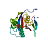
| ||||||||
|---|---|---|---|---|---|---|---|---|---|
| 1 |
| ||||||||
| Unit cell |
|
-Components
| #1: Protein | Mass: 18255.309 Da / Num. of mol.: 1 / Mutation: S32A Source method: isolated from a genetically manipulated source Source: (gene. exp.) Homo sapiens (human) / Gene: PIN1 / Plasmid: pET28b+ / Production host:  |
|---|---|
| #2: Chemical | ChemComp-1PG /   Mass: 252.305 Da / Num. of mol.: 1 / Source method: obtained synthetically / Formula: C11H24O6 Mass: 252.305 Da / Num. of mol.: 1 / Source method: obtained synthetically / Formula: C11H24O6 |
| #3: Chemical | ChemComp-SO4 /   Mass: 96.063 Da / Num. of mol.: 1 / Source method: obtained synthetically / Formula: SO4 Mass: 96.063 Da / Num. of mol.: 1 / Source method: obtained synthetically / Formula: SO4 |
| #4: Water | ChemComp-HOH /  Mass: 18.015 Da / Num. of mol.: 139 / Source method: isolated from a natural source / Formula: H2O Mass: 18.015 Da / Num. of mol.: 139 / Source method: isolated from a natural source / Formula: H2O |
-Experimental details
-Experiment
| Experiment | Method: X-RAY DIFFRACTION / Number of used crystals: 1 |
|---|
- Sample preparation
| Crystal | Density Matthews: 2.26 Å3/Da / Density % sol: 45.55 % |
|---|---|
| Crystal grow | Temperature: 278 K / Method: vapor diffusion, hanging drop / pH: 7.5 Details: 2.5M ammonium sulfate, 100mM HEPES-NA(pH7.5), 2% PEG400, 2mM DTT, VAPOR DIFFUSION, HANGING DROP, temperature 278K |
-Data collection
| Diffraction | Mean temperature: 298 K |
|---|---|
| Diffraction source | Source: ROTATING ANODE / Type: RIGAKU / Wavelength: 1.54178 Å |
| Detector | Type: RIGAKU RAXIS IV++ / Detector: IMAGE PLATE / Date: Nov 3, 2006 |
| Radiation | Monochromator: CN1707 / Protocol: SINGLE WAVELENGTH / Monochromatic (M) / Laue (L): M / Scattering type: x-ray |
| Radiation wavelength | Wavelength: 1.54178 Å / Relative weight: 1 |
| Reflection | Resolution: 1.8→50 Å / Num. all: 16318 / Num. obs: 15430 / % possible obs: 94.6 % / Observed criterion σ(F): 2 / Observed criterion σ(I): 2 / Rsym value: 0.062 / Net I/σ(I): 15 |
- Processing
| Software |
| ||||||||||||||||||||
|---|---|---|---|---|---|---|---|---|---|---|---|---|---|---|---|---|---|---|---|---|---|
| Refinement | Method to determine structure: MOLECULAR REPLACEMENT Starting model: PDB ENTRY 1PIN Resolution: 2→25 Å / Cross valid method: THROUGHOUT / σ(F): 2 / σ(I): 2 / Stereochemistry target values: Engh & Huber
| ||||||||||||||||||||
| Refinement step | Cycle: LAST / Resolution: 2→25 Å
| ||||||||||||||||||||
| Refine LS restraints |
|
