 Movie
Movie Controller


+ Open data

- Basic information
| Entry | Database: PDB / ID: 2z8w | ||||||
|---|---|---|---|---|---|---|---|
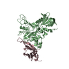
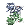
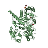
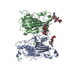
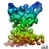
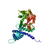
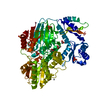
| Title | Structure of an IgNAR-AMA1 complex | ||||||
 Components Components |
| ||||||
 Keywords Keywords | IMMUNE SYSTEM / AMA1-VNAR complex / 14I1-M15 / Receptor | ||||||
| Function / homology |  Function and homology information Function and homology informationapical complex / microneme / host cell surface binding / symbiont entry into host / membrane Similarity search - Function | ||||||
| Biological species |   Orectolobus maculatus (spotted wobbegong) Orectolobus maculatus (spotted wobbegong) | ||||||
| Method |  X-RAY DIFFRACTION / SYNCHROTRON / MOLECULAR REPLACEMENT / Resolution: 2.45 Å X-RAY DIFFRACTION / SYNCHROTRON / MOLECULAR REPLACEMENT / Resolution: 2.45 Å | ||||||
 Authors Authors | Streltsov, V.A. / Henderson, K.A. / Batchelor, A.H. / Coley, A.M. / Nuttall, S.D. | ||||||
 Citation Citation | Journal: Structure / Year: 2007 Title: Structure of an IgNAR-AMA1 Complex: Targeting a Conserved Hydrophobic Cleft Broadens Malarial Strain Recognition Authors: Henderson, K.A. / Streltsov, V.A. / Coley, A.M. / Dolezal, O. / Hudson, P.J. / Batchelor, A.H. / Gupta, A. / Bai, T. / Murphy, V.J. / Anders, R.F. / Foley, M. / Nuttall, S.D. | ||||||
| History |
|
- Structure visualization
| Structure viewer | Molecule: MolmilJmol/JSmol |
|---|
- Downloads & links
-Download
| PDBx/mmCIF format | 2z8w.cif.gz | 198.1 KB | Display | PDBx/mmCIF format |
|---|---|---|---|---|
| PDB format | pdb2z8w.ent.gz | 157.3 KB | Display | PDB format |
| PDBx/mmJSON format | 2z8w.json.gz | Tree view | PDBx/mmJSON format | |
| Others |  Other downloads Other downloads |
-Validation report
| Arichive directory | https://data.pdbj.org/pub/pdb/validation_reports/z8/2z8wftp://data.pdbj.org/pub/pdb/validation_reports/z8/2z8w | HTTPS FTP |
|---|
-Related structure data
| Related structure data | 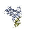 2z8vC  1verS 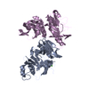 1z40S S: Starting model for refinement C: citing same article ( |
|---|---|
| Similar structure data |


-Links
 PDBj
PDBj- Assembly
| Deposited unit | 
| ||||||||
|---|---|---|---|---|---|---|---|---|---|
| 1 | 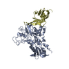
| ||||||||
| 2 | 
| ||||||||
| Unit cell |
|
-Components
| #1: Protein | Mass: 38360.047 Da / Num. of mol.: 2 / Fragment: domain I, II, UNP residues 104-438 Source method: isolated from a genetically manipulated source Source: (gene. exp.) Strain: 3D7 / Plasmid: pPROEXHTB / Species (production host): Escherichia coli / Production host:  #2: Protein | Mass: 12861.390 Da / Num. of mol.: 2 / Mutation: P90L/G92R Source method: isolated from a genetically manipulated source Source: (gene. exp.) Orectolobus maculatus (spotted wobbegong)Plasmid: pGC / Production host: #3: Water | ChemComp-HOH / |  Mass: 18.015 Da / Num. of mol.: 476 / Source method: isolated from a natural source / Formula: H2O Mass: 18.015 Da / Num. of mol.: 476 / Source method: isolated from a natural source / Formula: H2OHas protein modification | Y | |
|---|
-Experimental details
-Experiment
| Experiment | Method: X-RAY DIFFRACTION / Number of used crystals: 1 |
|---|
- Sample preparation
| Crystal | Density Matthews: 2.32 Å3/Da / Density % sol: 47.07 % |
|---|---|
| Crystal grow | Temperature: 298 K / Method: vapor diffusion, hanging drop / pH: 4.2 Details: 0.1M phosphate citrate pH 4.2, 0.2M NaCl, 20% PEG 8000, VAPOR DIFFUSION, HANGING DROP, temperature 298K |
-Data collection
| Diffraction | Mean temperature: 100 K |
|---|---|
| Diffraction source | Source: SYNCHROTRON / Site: Photon Factory  / Beamline: BL-18B / Wavelength: 0.96426 Å / Beamline: BL-18B / Wavelength: 0.96426 Å |
| Detector | Type: ADSC QUANTUM 4 / Detector: CCD / Date: Jun 21, 2006 / Details: mirrors |
| Radiation | Monochromator: Si(111) double crystal / Protocol: SINGLE WAVELENGTH / Monochromatic (M) / Laue (L): M / Scattering type: x-ray |
| Radiation wavelength | Wavelength: 0.96426 Å / Relative weight: 1 |
| Reflection | Resolution: 2.45→66.23 Å / Num. all: 28622 / Num. obs: 28622 / Observed criterion σ(F): 1 / Observed criterion σ(I): 1 / Redundancy: 5 % / Rmerge(I) obs: 0.13 / Net I/σ(I): 14.4 |
| Reflection shell | Resolution: 2.45→2.51 Å / Redundancy: 2.1 % / Rmerge(I) obs: 0.57 / Mean I/σ(I) obs: 1.6 |
- Processing
| Software |
| ||||||||||||||||||||||||||||||||||||||||||||||||||||||||||||||||||||||||||||||||||||||||||||||||||||||||||||||||||||||||||||||||||||||||||||||||||||||||||||||||||||||||||
|---|---|---|---|---|---|---|---|---|---|---|---|---|---|---|---|---|---|---|---|---|---|---|---|---|---|---|---|---|---|---|---|---|---|---|---|---|---|---|---|---|---|---|---|---|---|---|---|---|---|---|---|---|---|---|---|---|---|---|---|---|---|---|---|---|---|---|---|---|---|---|---|---|---|---|---|---|---|---|---|---|---|---|---|---|---|---|---|---|---|---|---|---|---|---|---|---|---|---|---|---|---|---|---|---|---|---|---|---|---|---|---|---|---|---|---|---|---|---|---|---|---|---|---|---|---|---|---|---|---|---|---|---|---|---|---|---|---|---|---|---|---|---|---|---|---|---|---|---|---|---|---|---|---|---|---|---|---|---|---|---|---|---|---|---|---|---|---|---|---|---|---|
| Refinement | Method to determine structure: MOLECULAR REPLACEMENT Starting model: PDB ENTRIES 1Z40 and 1VER Resolution: 2.45→38.33 Å / Cor.coef. Fo:Fc: 0.939 / Cor.coef. Fo:Fc free: 0.865 / SU B: 23.776 / SU ML: 0.289 / TLS residual ADP flag: LIKELY RESIDUAL / Cross valid method: THROUGHOUT / σ(F): 1 / σ(I): 1 / ESU R Free: 0.379 / Stereochemistry target values: MAXIMUM LIKELIHOOD / Details: HYDROGENS HAVE BEEN ADDED IN THE RIDING POSITIONS
| ||||||||||||||||||||||||||||||||||||||||||||||||||||||||||||||||||||||||||||||||||||||||||||||||||||||||||||||||||||||||||||||||||||||||||||||||||||||||||||||||||||||||||
| Solvent computation | Ion probe radii: 0.8 Å / Shrinkage radii: 0.8 Å / VDW probe radii: 1.4 Å / Solvent model: BABINET MODEL WITH MASK | ||||||||||||||||||||||||||||||||||||||||||||||||||||||||||||||||||||||||||||||||||||||||||||||||||||||||||||||||||||||||||||||||||||||||||||||||||||||||||||||||||||||||||
| Displacement parameters | Biso mean: 57.778 Å2
| ||||||||||||||||||||||||||||||||||||||||||||||||||||||||||||||||||||||||||||||||||||||||||||||||||||||||||||||||||||||||||||||||||||||||||||||||||||||||||||||||||||||||||
| Refinement step | Cycle: LAST / Resolution: 2.45→38.33 Å
| ||||||||||||||||||||||||||||||||||||||||||||||||||||||||||||||||||||||||||||||||||||||||||||||||||||||||||||||||||||||||||||||||||||||||||||||||||||||||||||||||||||||||||
| Refine LS restraints |
| ||||||||||||||||||||||||||||||||||||||||||||||||||||||||||||||||||||||||||||||||||||||||||||||||||||||||||||||||||||||||||||||||||||||||||||||||||||||||||||||||||||||||||
| LS refinement shell | Resolution: 2.45→2.514 Å / Total num. of bins used: 20
| ||||||||||||||||||||||||||||||||||||||||||||||||||||||||||||||||||||||||||||||||||||||||||||||||||||||||||||||||||||||||||||||||||||||||||||||||||||||||||||||||||||||||||
| Refinement TLS params. | Method: refined / Refine-ID: X-RAY DIFFRACTION
| ||||||||||||||||||||||||||||||||||||||||||||||||||||||||||||||||||||||||||||||||||||||||||||||||||||||||||||||||||||||||||||||||||||||||||||||||||||||||||||||||||||||||||
| Refinement TLS group |
|
