 ムービー
ムービー コントローラー


+ データを開く

- 基本情報
基本情報
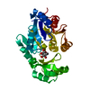
| 登録情報 | データベース: PDB / ID: 2z6c | ||||||
|---|---|---|---|---|---|---|---|
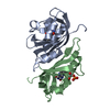
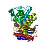
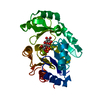
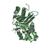
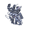
| タイトル | Crystal structure of LOV1 domain of phototropin1 from Arabidopsis thaliana | ||||||
 要素 要素 | Phototropin-1 | ||||||
 キーワード キーワード | TRANSFERASE / PAS-FOLD / LOV-FOLD / ATP-binding / Chromophore / Cytoplasm / Flavoprotein / FMN / Kinase / Membrane / Nucleotide-binding / Phosphorylation / Photoreceptor protein / Receptor / Sensory transduction / Serine/threonine-protein kinase | ||||||
| 機能・相同性 |  機能・相同性情報 機能・相同性情報chloroplast avoidance movement / chloroplast accumulation movement / cellular response to blue light / regulation of proton transport / phototropism / regulation of stomatal movement / blue light photoreceptor activity / response to blue light / plant-type vacuole / circadian rhythm ...chloroplast avoidance movement / chloroplast accumulation movement / cellular response to blue light / regulation of proton transport / phototropism / regulation of stomatal movement / blue light photoreceptor activity / response to blue light / plant-type vacuole / circadian rhythm / cytoplasmic side of plasma membrane / kinase activity / FMN binding / protein autophosphorylation / non-specific serine/threonine protein kinase / protein kinase activity / protein serine kinase activity / protein serine/threonine kinase activity / mRNA binding / cell surface / ATP binding / identical protein binding / cytoplasm 類似検索 - 分子機能 | ||||||
| 生物種 |  | ||||||
| 手法 |  X線回折 / シンクロトロン / 分子置換 / 解像度: 2.1 Å X線回折 / シンクロトロン / 分子置換 / 解像度: 2.1 Å | ||||||
 データ登録者 データ登録者 | Nakasako, M. / Matsuoka, D. / Tokutomi, S. | ||||||
 引用 引用 | ジャーナル: J.Mol.Biol. / 年: 2008 タイトル: Structural basis of the LOV1 dimerization of Arabidopsis phototropins 1 and 2 著者: Nakasako, M. / Zikihara, K. / Matsuoka, D. / Katsura, H. / Tokutomi, S. #1: ジャーナル: Acta Crystallogr.,Sect.F / 年: 2008 タイトル: Crystallization and preliminary X-ray diffraction analysis of the LOV1 domains of phototropin 1 and 2 from Arabidopsis thaliana 著者: Nakasako, M. / Hirata, M. / Shimizu, N. / Hosokawa, S. / Matsuoka, D. / Oka, T. / Yamamoto, M. / Tokutomi, S. #2: ジャーナル: Biochemistry / 年: 2004 タイトル: Light-induced structural changes of LOV domain-containing polypeptides from Arabidopsis phototropin 1 and 2 studied by small-angle X-ray scattering 著者: Nakasako, M. / Iwata, T. / Matsuoka, D. / Tokutomi, S. #3: ジャーナル: FEBS Lett. / 年: 2005 タイトル: Quaternary structure of LOV-domain containing polypeptide of Arabidopsis FKF1 protein 著者: Nakasako, M. / Matsuoka, D. / Zikihara, K. / Tokutomi, S. | ||||||
| 履歴 |
|
- 構造の表示
構造の表示
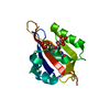
| 構造ビューア | 分子: MolmilJmol/JSmol |
|---|
- ダウンロードとリンク
-ダウンロード
| PDBx/mmCIF形式 | 2z6c.cif.gz | 65.5 KB | 表示 | PDBx/mmCIF形式 |
|---|---|---|---|---|
| PDB形式 | pdb2z6c.ent.gz | 48.3 KB | 表示 | PDB形式 |
| PDBx/mmJSON形式 | 2z6c.json.gz | ツリー表示 | PDBx/mmJSON形式 | |
| その他 |  その他のダウンロード その他のダウンロード |
-検証レポート
| 文書・要旨 | 2z6c_validation.pdf.gz | 1 MB | 表示 | wwPDB検証レポート |
|---|---|---|---|---|
| 文書・詳細版 | 2z6c_full_validation.pdf.gz | 1 MB | 表示 | |
| XML形式データ | 2z6c_validation.xml.gz | 14 KB | 表示 | |
| CIF形式データ | 2z6c_validation.cif.gz | 18.8 KB | 表示 | |
| アーカイブディレクトリ | https://data.pdbj.org/pub/pdb/validation_reports/z6/2z6cftp://data.pdbj.org/pub/pdb/validation_reports/z6/2z6c | HTTPS FTP |
-関連構造データ


-リンク
 PDBj
PDBj


- 集合体
集合体
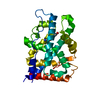
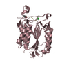
| 登録構造単位 | 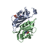
| ||||||||
|---|---|---|---|---|---|---|---|---|---|
| 1 |
| ||||||||
| 単位格子 |
|
-要素
| #1: タンパク質 | 分子量: 14199.078 Da / 分子数: 2 / 断片: UNP residues 180-308, LOV1 domain / 由来タイプ: 組換発現 由来: (組換発現) 遺伝子: PHOT1, JK224, NPH1, RPT1 / 発現宿主:  参照: UniProt: O48963, non-specific serine/threonine protein kinase #2: 化合物 |   分子量: 456.344 Da / 分子数: 2 / 由来タイプ: 合成 / 式: C17H21N4O9P 分子量: 456.344 Da / 分子数: 2 / 由来タイプ: 合成 / 式: C17H21N4O9P#3: 水 | ChemComp-HOH / |  分子量: 18.015 Da / 分子数: 140 / 由来タイプ: 天然 / 式: H2O 分子量: 18.015 Da / 分子数: 140 / 由来タイプ: 天然 / 式: H2OHas protein modification | Y | |
|---|
-実験情報
-実験
| 実験 | 手法: X線回折 / 使用した結晶の数: 1 |
|---|
- 試料調製
試料調製
| 結晶 | マシュー密度: 2.47 Å3/Da / 溶媒含有率: 50.3 % |
|---|---|
| 結晶化 | 温度: 293 K / 手法: 蒸気拡散法, ハンギングドロップ法 / pH: 8 詳細: ammonium sulfate, pH8, VAPOR DIFFUSION, HANGING DROP, temperature 293K |
-データ収集
| 放射光源 | 由来: シンクロトロン / サイト: SPring-8  / ビームライン: BL44B2 / 波長: 1 Å / ビームライン: BL44B2 / 波長: 1 Å |
|---|---|
| 検出器 | タイプ: ADSC QUANTUM 210 / 検出器: CCD / 日付: 2007年6月22日 / 詳細: monochrometer/focusing mirror |
| 放射 | モノクロメーター: Si / プロトコル: SINGLE WAVELENGTH / 単色(M)・ラウエ(L): M / 散乱光タイプ: x-ray |
| 放射波長 | 波長: 1 Å / 相対比: 1 |
| 反射 | 解像度: 2.1→50 Å / Num. obs: 16967 / % possible obs: 99.9 % / Observed criterion σ(I): 1 / 冗長度: 13.9 % / Rmerge(I) obs: 0.057 / Net I/σ(I): 41.8 |
| 反射 シェル | 解像度: 2.1→2.17 Å / Rmerge(I) obs: 0.352 / Mean I/σ(I) obs: 4.8 / % possible all: 99.2 |
- 解析
解析
| ソフトウェア |
| ||||||||||||||||||||||||||||||||||||||||||||||||||||||||||||||||||||||||||||||||||||||||||||||||||||||||||||||||||||||||||||||||||||||||||||||||||||||||||||||||||||||||||
|---|---|---|---|---|---|---|---|---|---|---|---|---|---|---|---|---|---|---|---|---|---|---|---|---|---|---|---|---|---|---|---|---|---|---|---|---|---|---|---|---|---|---|---|---|---|---|---|---|---|---|---|---|---|---|---|---|---|---|---|---|---|---|---|---|---|---|---|---|---|---|---|---|---|---|---|---|---|---|---|---|---|---|---|---|---|---|---|---|---|---|---|---|---|---|---|---|---|---|---|---|---|---|---|---|---|---|---|---|---|---|---|---|---|---|---|---|---|---|---|---|---|---|---|---|---|---|---|---|---|---|---|---|---|---|---|---|---|---|---|---|---|---|---|---|---|---|---|---|---|---|---|---|---|---|---|---|---|---|---|---|---|---|---|---|---|---|---|---|---|---|---|
| 精密化 | 構造決定の手法: 分子置換 開始モデル: PDB ENTRY 1N9L 解像度: 2.1→15 Å / Cor.coef. Fo:Fc: 0.949 / Cor.coef. Fo:Fc free: 0.92 / SU B: 4.128 / SU ML: 0.115 / 交差検証法: THROUGHOUT / ESU R: 0.211 / ESU R Free: 0.187 / 立体化学のターゲット値: MAXIMUM LIKELIHOOD
| ||||||||||||||||||||||||||||||||||||||||||||||||||||||||||||||||||||||||||||||||||||||||||||||||||||||||||||||||||||||||||||||||||||||||||||||||||||||||||||||||||||||||||
| 溶媒の処理 | イオンプローブ半径: 0.8 Å / 減衰半径: 0.8 Å / VDWプローブ半径: 1.4 Å / 溶媒モデル: MASK | ||||||||||||||||||||||||||||||||||||||||||||||||||||||||||||||||||||||||||||||||||||||||||||||||||||||||||||||||||||||||||||||||||||||||||||||||||||||||||||||||||||||||||
| 原子変位パラメータ | Biso mean: 29.54 Å2
| ||||||||||||||||||||||||||||||||||||||||||||||||||||||||||||||||||||||||||||||||||||||||||||||||||||||||||||||||||||||||||||||||||||||||||||||||||||||||||||||||||||||||||
| 精密化ステップ | サイクル: LAST / 解像度: 2.1→15 Å
| ||||||||||||||||||||||||||||||||||||||||||||||||||||||||||||||||||||||||||||||||||||||||||||||||||||||||||||||||||||||||||||||||||||||||||||||||||||||||||||||||||||||||||
| 拘束条件 |
| ||||||||||||||||||||||||||||||||||||||||||||||||||||||||||||||||||||||||||||||||||||||||||||||||||||||||||||||||||||||||||||||||||||||||||||||||||||||||||||||||||||||||||
| LS精密化 シェル | 解像度: 2.1→2.16 Å / Total num. of bins used: 20
|
