 Movie
Movie Controller

[English] 日本語
 Yorodumi
- PDB-2z6c: Crystal structure of LOV1 domain of phototropin1 from Arabidopsis... -
+ Open data

- Basic information
| Entry | Database: PDB / ID: 2z6c | ||||||
|---|---|---|---|---|---|---|---|
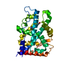
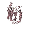
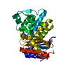
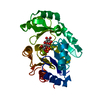
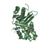
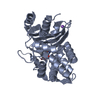
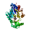
| Title | Crystal structure of LOV1 domain of phototropin1 from Arabidopsis thaliana | ||||||
 Components Components | Phototropin-1 | ||||||
 Keywords Keywords | TRANSFERASE / PAS-FOLD / LOV-FOLD / ATP-binding / Chromophore / Cytoplasm / Flavoprotein / FMN / Kinase / Membrane / Nucleotide-binding / Phosphorylation / Photoreceptor protein / Receptor / Sensory transduction / Serine/threonine-protein kinase | ||||||
| Function / homology |  Function and homology information Function and homology informationchloroplast avoidance movement / chloroplast accumulation movement / cellular response to blue light / regulation of proton transport / leaf phyllotactic patterning / response to red or far red light / regulation of leaf morphogenesis / phototropism / blue light signaling pathway / regulation of stomatal movement ...chloroplast avoidance movement / chloroplast accumulation movement / cellular response to blue light / regulation of proton transport / leaf phyllotactic patterning / response to red or far red light / regulation of leaf morphogenesis / phototropism / blue light signaling pathway / regulation of stomatal movement / blue light photoreceptor activity / response to blue light / plant-type vacuole / circadian rhythm / cytoplasmic side of plasma membrane / kinase activity / FMN binding / protein autophosphorylation / protein kinase activity / non-specific serine/threonine protein kinase / protein serine kinase activity / protein serine/threonine kinase activity / mRNA binding / cell surface / ATP binding / identical protein binding / nucleus / plasma membrane / cytoplasm Similarity search - Function | ||||||
| Biological species |  | ||||||
| Method |  X-RAY DIFFRACTION / SYNCHROTRON / MOLECULAR REPLACEMENT / Resolution: 2.1 Å X-RAY DIFFRACTION / SYNCHROTRON / MOLECULAR REPLACEMENT / Resolution: 2.1 Å | ||||||
 Authors Authors | Nakasako, M. / Matsuoka, D. / Tokutomi, S. | ||||||
 Citation Citation | Journal: J.Mol.Biol. / Year: 2008 Title: Structural basis of the LOV1 dimerization of Arabidopsis phototropins 1 and 2 Authors: Nakasako, M. / Zikihara, K. / Matsuoka, D. / Katsura, H. / Tokutomi, S. #1: Journal: Acta Crystallogr.,Sect.F / Year: 2008 Title: Crystallization and preliminary X-ray diffraction analysis of the LOV1 domains of phototropin 1 and 2 from Arabidopsis thaliana Authors: Nakasako, M. / Hirata, M. / Shimizu, N. / Hosokawa, S. / Matsuoka, D. / Oka, T. / Yamamoto, M. / Tokutomi, S. #2: Journal: Biochemistry / Year: 2004 Title: Light-induced structural changes of LOV domain-containing polypeptides from Arabidopsis phototropin 1 and 2 studied by small-angle X-ray scattering Authors: Nakasako, M. / Iwata, T. / Matsuoka, D. / Tokutomi, S. #3: Journal: FEBS Lett. / Year: 2005 Title: Quaternary structure of LOV-domain containing polypeptide of Arabidopsis FKF1 protein Authors: Nakasako, M. / Matsuoka, D. / Zikihara, K. / Tokutomi, S. | ||||||
| History |
|
- Structure visualization
| Structure viewer | Molecule: MolmilJmol/JSmol |
|---|
- Downloads & links
-Download
| PDBx/mmCIF format | 2z6c.cif.gz | 65.5 KB | Display | PDBx/mmCIF format |
|---|---|---|---|---|
| PDB format | pdb2z6c.ent.gz | 48.3 KB | Display | PDB format |
| PDBx/mmJSON format | 2z6c.json.gz | Tree view | PDBx/mmJSON format | |
| Others |  Other downloads Other downloads |
-Validation report
| Arichive directory | https://data.pdbj.org/pub/pdb/validation_reports/z6/2z6cftp://data.pdbj.org/pub/pdb/validation_reports/z6/2z6c | HTTPS FTP |
|---|
-Related structure data
| Related structure data | 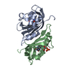 2z6dC 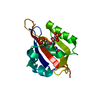 1n9lS S: Starting model for refinement C: citing same article ( |
|---|---|
| Similar structure data |


-Links
 PDBj
PDBj


- Assembly
| Deposited unit | 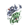
| ||||||||
|---|---|---|---|---|---|---|---|---|---|
| 1 |
| ||||||||
| Unit cell |
|
-Components
| #1: Protein | Mass: 14199.078 Da / Num. of mol.: 2 / Fragment: UNP residues 180-308, LOV1 domain Source method: isolated from a genetically manipulated source Source: (gene. exp.)  References: UniProt: O48963, non-specific serine/threonine protein kinase #2: Chemical |   Mass: 456.344 Da / Num. of mol.: 2 / Source method: obtained synthetically / Formula: C17H21N4O9P Mass: 456.344 Da / Num. of mol.: 2 / Source method: obtained synthetically / Formula: C17H21N4O9P#3: Water | ChemComp-HOH / |  Mass: 18.015 Da / Num. of mol.: 140 / Source method: isolated from a natural source / Formula: H2O Mass: 18.015 Da / Num. of mol.: 140 / Source method: isolated from a natural source / Formula: H2OHas protein modification | Y | |
|---|
-Experimental details
-Experiment
| Experiment | Method: X-RAY DIFFRACTION / Number of used crystals: 1 |
|---|
- Sample preparation
| Crystal | Density Matthews: 2.47 Å3/Da / Density % sol: 50.3 % |
|---|---|
| Crystal grow | Temperature: 293 K / Method: vapor diffusion, hanging drop / pH: 8 Details: ammonium sulfate, pH8, VAPOR DIFFUSION, HANGING DROP, temperature 293K |
-Data collection
| Diffraction source | Source: SYNCHROTRON / Site: SPring-8  / Beamline: BL44B2 / Wavelength: 1 Å / Beamline: BL44B2 / Wavelength: 1 Å |
|---|---|
| Detector | Type: ADSC QUANTUM 210 / Detector: CCD / Date: Jun 22, 2007 / Details: monochrometer/focusing mirror |
| Radiation | Monochromator: Si / Protocol: SINGLE WAVELENGTH / Monochromatic (M) / Laue (L): M / Scattering type: x-ray |
| Radiation wavelength | Wavelength: 1 Å / Relative weight: 1 |
| Reflection | Resolution: 2.1→50 Å / Num. obs: 16967 / % possible obs: 99.9 % / Observed criterion σ(I): 1 / Redundancy: 13.9 % / Rmerge(I) obs: 0.057 / Net I/σ(I): 41.8 |
| Reflection shell | Resolution: 2.1→2.17 Å / Rmerge(I) obs: 0.352 / Mean I/σ(I) obs: 4.8 / % possible all: 99.2 |
- Processing
| Software |
| ||||||||||||||||||||||||||||||||||||||||||||||||||||||||||||||||||||||||||||||||||||||||||||||||||||||||||||||||||||||||||||||||||||||||||||||||||||||||||||||||||||||||||
|---|---|---|---|---|---|---|---|---|---|---|---|---|---|---|---|---|---|---|---|---|---|---|---|---|---|---|---|---|---|---|---|---|---|---|---|---|---|---|---|---|---|---|---|---|---|---|---|---|---|---|---|---|---|---|---|---|---|---|---|---|---|---|---|---|---|---|---|---|---|---|---|---|---|---|---|---|---|---|---|---|---|---|---|---|---|---|---|---|---|---|---|---|---|---|---|---|---|---|---|---|---|---|---|---|---|---|---|---|---|---|---|---|---|---|---|---|---|---|---|---|---|---|---|---|---|---|---|---|---|---|---|---|---|---|---|---|---|---|---|---|---|---|---|---|---|---|---|---|---|---|---|---|---|---|---|---|---|---|---|---|---|---|---|---|---|---|---|---|---|---|---|
| Refinement | Method to determine structure: MOLECULAR REPLACEMENT Starting model: PDB ENTRY 1N9L Resolution: 2.1→15 Å / Cor.coef. Fo:Fc: 0.949 / Cor.coef. Fo:Fc free: 0.92 / SU B: 4.128 / SU ML: 0.115 / Cross valid method: THROUGHOUT / ESU R: 0.211 / ESU R Free: 0.187 / Stereochemistry target values: MAXIMUM LIKELIHOOD
| ||||||||||||||||||||||||||||||||||||||||||||||||||||||||||||||||||||||||||||||||||||||||||||||||||||||||||||||||||||||||||||||||||||||||||||||||||||||||||||||||||||||||||
| Solvent computation | Ion probe radii: 0.8 Å / Shrinkage radii: 0.8 Å / VDW probe radii: 1.4 Å / Solvent model: MASK | ||||||||||||||||||||||||||||||||||||||||||||||||||||||||||||||||||||||||||||||||||||||||||||||||||||||||||||||||||||||||||||||||||||||||||||||||||||||||||||||||||||||||||
| Displacement parameters | Biso mean: 29.54 Å2
| ||||||||||||||||||||||||||||||||||||||||||||||||||||||||||||||||||||||||||||||||||||||||||||||||||||||||||||||||||||||||||||||||||||||||||||||||||||||||||||||||||||||||||
| Refinement step | Cycle: LAST / Resolution: 2.1→15 Å
| ||||||||||||||||||||||||||||||||||||||||||||||||||||||||||||||||||||||||||||||||||||||||||||||||||||||||||||||||||||||||||||||||||||||||||||||||||||||||||||||||||||||||||
| Refine LS restraints |
| ||||||||||||||||||||||||||||||||||||||||||||||||||||||||||||||||||||||||||||||||||||||||||||||||||||||||||||||||||||||||||||||||||||||||||||||||||||||||||||||||||||||||||
| LS refinement shell | Resolution: 2.1→2.16 Å / Total num. of bins used: 20
|
