 Movie
Movie Controller

[English] 日本語
 Yorodumi
- PDB-2z3h: Crystal structure of blasticidin S deaminase (BSD) complexed with... -
+ Open data

- Basic information
| Entry | Database: PDB / ID: 2z3h | ||||||
|---|---|---|---|---|---|---|---|
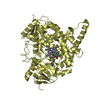
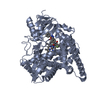
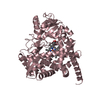
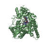
| Title | Crystal structure of blasticidin S deaminase (BSD) complexed with deaminohydroxy blasticidin S | ||||||
 Components Components | Blasticidin-S deaminase | ||||||
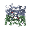
 Keywords Keywords | HYDROLASE / CYTIDINE DEAMINASE FAMILY / ZINC / TETRAMER | ||||||
| Function / homology |  Function and homology information Function and homology informationblasticidin-S deaminase / blasticidin-S deaminase activity / pyrimidine-containing compound metabolic process / nucleobase-containing small molecule metabolic process / cytidine deaminase activity / response to antibiotic / zinc ion binding / identical protein binding / cytosol Similarity search - Function | ||||||
| Biological species |  | ||||||
| Method |  X-RAY DIFFRACTION / SYNCHROTRON / MOLECULAR REPLACEMENT / Resolution: 1.5 Å X-RAY DIFFRACTION / SYNCHROTRON / MOLECULAR REPLACEMENT / Resolution: 1.5 Å | ||||||
 Authors Authors | Kumasaka, T. / Yamamoto, M. / Furuichi, M. / Nakasako, M. / Kimura, M. / Yamaguchi, I. / Ueki, T. | ||||||
 Citation Citation | Journal: J.Biol.Chem. / Year: 2007 Title: Crystal structures of blasticidin S deaminase (BSD): implications for dynamic properties of catalytic zinc Authors: Kumasaka, T. / Yamamoto, M. / Furuichi, M. / Nakasako, M. / Teh, A.H. / Kimura, M. / Yamaguchi, I. / Ueki, T. #1: Journal: Acta Crystallogr.,Sect.D / Year: 1999 Title: Crystallization and Preliminary X-Ray Diffraction Studies of Blasticidin S Deaminase from Aspergillus Terreus Authors: Nakasako, M. / Kimura, M. / Yamaguchi, I. | ||||||
| History |
|
- Structure visualization
| Structure viewer | Molecule: MolmilJmol/JSmol |
|---|
- Downloads & links
-Download
| PDBx/mmCIF format | 2z3h.cif.gz | 123.8 KB | Display | PDBx/mmCIF format |
|---|---|---|---|---|
| PDB format | pdb2z3h.ent.gz | 95.7 KB | Display | PDB format |
| PDBx/mmJSON format | 2z3h.json.gz | Tree view | PDBx/mmJSON format | |
| Others |  Other downloads Other downloads |
-Validation report
| Arichive directory | https://data.pdbj.org/pub/pdb/validation_reports/z3/2z3hftp://data.pdbj.org/pub/pdb/validation_reports/z3/2z3h | HTTPS FTP |
|---|
-Related structure data
| Related structure data |  1wn5SC  1wn6C  2z3gC  2z3iC  2z3jC C: citing same article ( S: Starting model for refinement |
|---|---|
| Similar structure data |

-Links
 PDBj
PDBj- Assembly
| Deposited unit | 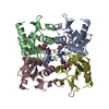
| ||||||||
|---|---|---|---|---|---|---|---|---|---|
| 1 |
| ||||||||
| Unit cell |
|
-Components
| #1: Protein | Mass: 13478.245 Da / Num. of mol.: 4 Source method: isolated from a genetically manipulated source Source: (gene. exp.)  #2: Chemical | ChemComp-ZN /   Mass: 65.409 Da / Num. of mol.: 4 / Source method: obtained synthetically / Formula: Zn Mass: 65.409 Da / Num. of mol.: 4 / Source method: obtained synthetically / Formula: Zn#3: Chemical | ChemComp-BLO /   Mass: 423.424 Da / Num. of mol.: 4 / Source method: obtained synthetically / Formula: C17H25N7O6 Mass: 423.424 Da / Num. of mol.: 4 / Source method: obtained synthetically / Formula: C17H25N7O6#4: Water | ChemComp-HOH / |  Mass: 18.015 Da / Num. of mol.: 682 / Source method: isolated from a natural source / Formula: H2O Mass: 18.015 Da / Num. of mol.: 682 / Source method: isolated from a natural source / Formula: H2O |
|---|
-Experimental details
-Experiment
| Experiment | Method: X-RAY DIFFRACTION / Number of used crystals: 1 |
|---|
- Sample preparation
| Crystal | Density Matthews: 2.55 Å3/Da / Density % sol: 51.85 % |
|---|---|
| Crystal grow | Temperature: 293 K / Method: vapor diffusion, hanging drop / pH: 7 Details: 20% PEG8000, 0.1M SODIUM CACODYLATE, 50MM MAGNESIUM CHLORIDE, pH 7.00, VAPOR DIFFUSION, HANGING DROP, temperature 293K |
-Data collection
| Diffraction | Mean temperature: 100 K |
|---|---|
| Diffraction source | Source: SYNCHROTRON / Site: SPring-8  / Beamline: BL45XU / Wavelength: 1 / Beamline: BL45XU / Wavelength: 1 |
| Detector | Type: RIGAKU RAXIS IV / Detector: IMAGE PLATE |
| Radiation | Monochromator: DIAMOND (400) / Protocol: SINGLE WAVELENGTH / Monochromatic (M) / Laue (L): M / Scattering type: x-ray |
| Radiation wavelength | Wavelength: 1 Å / Relative weight: 1 |
| Reflection | Resolution: 1.5→50 Å / Num. obs: 89147 / % possible obs: 99.8 % / Observed criterion σ(I): 0 / Rmerge(I) obs: 0.091 |
- Processing
| Software |
| ||||||||||||||||||||||||||||||||||||||||||||||||||||||||||||||||||||||||||||||||||||||||||||||||||||||||||||||||||||||||||||||||||||||||||||||||||||||||||||||||||||||||||
|---|---|---|---|---|---|---|---|---|---|---|---|---|---|---|---|---|---|---|---|---|---|---|---|---|---|---|---|---|---|---|---|---|---|---|---|---|---|---|---|---|---|---|---|---|---|---|---|---|---|---|---|---|---|---|---|---|---|---|---|---|---|---|---|---|---|---|---|---|---|---|---|---|---|---|---|---|---|---|---|---|---|---|---|---|---|---|---|---|---|---|---|---|---|---|---|---|---|---|---|---|---|---|---|---|---|---|---|---|---|---|---|---|---|---|---|---|---|---|---|---|---|---|---|---|---|---|---|---|---|---|---|---|---|---|---|---|---|---|---|---|---|---|---|---|---|---|---|---|---|---|---|---|---|---|---|---|---|---|---|---|---|---|---|---|---|---|---|---|---|---|---|
| Refinement | Method to determine structure: MOLECULAR REPLACEMENT Starting model: PDB ENTRY 1WN5 Resolution: 1.5→50 Å / Cor.coef. Fo:Fc: 0.96 / Cor.coef. Fo:Fc free: 0.957 / SU B: 0.985 / SU ML: 0.038 / Cross valid method: THROUGHOUT / σ(F): 0 / ESU R: 0.068 / ESU R Free: 0.065 / Stereochemistry target values: ENGH & HUBER
| ||||||||||||||||||||||||||||||||||||||||||||||||||||||||||||||||||||||||||||||||||||||||||||||||||||||||||||||||||||||||||||||||||||||||||||||||||||||||||||||||||||||||||
| Solvent computation | Ion probe radii: 0.8 Å / Shrinkage radii: 0.8 Å / VDW probe radii: 1.2 Å / Solvent model: MASK | ||||||||||||||||||||||||||||||||||||||||||||||||||||||||||||||||||||||||||||||||||||||||||||||||||||||||||||||||||||||||||||||||||||||||||||||||||||||||||||||||||||||||||
| Displacement parameters | Biso mean: 14.88 Å2
| ||||||||||||||||||||||||||||||||||||||||||||||||||||||||||||||||||||||||||||||||||||||||||||||||||||||||||||||||||||||||||||||||||||||||||||||||||||||||||||||||||||||||||
| Refinement step | Cycle: LAST / Resolution: 1.5→50 Å
| ||||||||||||||||||||||||||||||||||||||||||||||||||||||||||||||||||||||||||||||||||||||||||||||||||||||||||||||||||||||||||||||||||||||||||||||||||||||||||||||||||||||||||
| Refine LS restraints |
| ||||||||||||||||||||||||||||||||||||||||||||||||||||||||||||||||||||||||||||||||||||||||||||||||||||||||||||||||||||||||||||||||||||||||||||||||||||||||||||||||||||||||||
| LS refinement shell | Resolution: 1.5→1.54 Å / Total num. of bins used: 20
|
