 Movie
Movie Controller


+ Open data

- Basic information
| Entry | Database: PDB / ID: 2yxt | ||||||
|---|---|---|---|---|---|---|---|
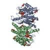
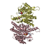
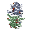
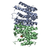
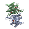
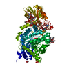
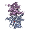
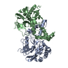
| Title | Human Pyridoxal Kinase | ||||||
 Components Components | Pyridoxal kinase | ||||||
 Keywords Keywords | TRANSFERASE / beta sheet with alpha helix / metal ion | ||||||
| Function / homology |  Function and homology information Function and homology informationpyridoxal metabolic process / pyridoxamine metabolic process / Vitamin B6 activation to pyridoxal phosphate / lithium ion binding / pyridoxal 5'-phosphate salvage / pyridoxal kinase / pyridoxal kinase activity / sodium ion binding / potassium ion binding / specific granule lumen ...pyridoxal metabolic process / pyridoxamine metabolic process / Vitamin B6 activation to pyridoxal phosphate / lithium ion binding / pyridoxal 5'-phosphate salvage / pyridoxal kinase / pyridoxal kinase activity / sodium ion binding / potassium ion binding / specific granule lumen / pyridoxal phosphate binding / secretory granule lumen / Neutrophil degranulation / magnesium ion binding / protein homodimerization activity / extracellular exosome / extracellular region / zinc ion binding / nucleoplasm / ATP binding / nucleus / cytosol Similarity search - Function | ||||||
| Biological species |  Homo sapiens (human) Homo sapiens (human) | ||||||
| Method |  X-RAY DIFFRACTION / MOLECULAR REPLACEMENT / Resolution: 2 Å X-RAY DIFFRACTION / MOLECULAR REPLACEMENT / Resolution: 2 Å | ||||||
 Authors Authors | Safo, M.K. / Musayev, F.N. / Ko, T.P. / Schirch, V. | ||||||
 Citation Citation | Journal: Protein Sci. / Year: 2007 Title: Crystal Structure of human pyridoxal kinase: structural basis of M(+) and M(2+) activation. Authors: Musayev, F.N. / di Salvo, M.L. / Ko, T.P. / Gandhi, A.K. / Goswami, A. / Schirch, V. / Safo, M.K. | ||||||
| History |
|
- Structure visualization
| Structure viewer | Molecule: MolmilJmol/JSmol |
|---|
- Downloads & links
-Download
| PDBx/mmCIF format | 2yxt.cif.gz | 147.6 KB | Display | PDBx/mmCIF format |
|---|---|---|---|---|
| PDB format | pdb2yxt.ent.gz | 115.5 KB | Display | PDB format |
| PDBx/mmJSON format | 2yxt.json.gz | Tree view | PDBx/mmJSON format | |
| Others |  Other downloads Other downloads |
-Validation report
| Arichive directory | https://data.pdbj.org/pub/pdb/validation_reports/yx/2yxtftp://data.pdbj.org/pub/pdb/validation_reports/yx/2yxt | HTTPS FTP |
|---|
-Related structure data
| Related structure data | 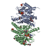 2yxuC 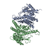 1lhpS S: Starting model for refinement C: citing same article ( |
|---|---|
| Similar structure data |


-Links
 PDBj
PDBj- Assembly
| Deposited unit | 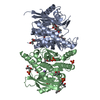
| |||||||||||||||
|---|---|---|---|---|---|---|---|---|---|---|---|---|---|---|---|---|
| 1 |
| |||||||||||||||
| 2 | 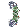
| |||||||||||||||
| Unit cell |
| |||||||||||||||
| Components on special symmetry positions |
|
-Components
| #1: Protein | Mass: 35143.266 Da / Num. of mol.: 2 Source method: isolated from a genetically manipulated source Source: (gene. exp.) Homo sapiens (human) / Gene: PDXK, PKH, PNK / Plasmid: pET 22b / Production host:  #2: Chemical |   Mass: 22.990 Da / Num. of mol.: 2 / Source method: obtained synthetically / Formula: Na Mass: 22.990 Da / Num. of mol.: 2 / Source method: obtained synthetically / Formula: Na#3: Chemical | ChemComp-PO4 /   Mass: 94.971 Da / Num. of mol.: 6 / Source method: obtained synthetically / Formula: PO4 Mass: 94.971 Da / Num. of mol.: 6 / Source method: obtained synthetically / Formula: PO4#4: Chemical | ChemComp-MPD / (   Mass: 118.174 Da / Num. of mol.: 14 / Source method: obtained synthetically / Formula: C6H14O2 / Comment: precipitant*YM Mass: 118.174 Da / Num. of mol.: 14 / Source method: obtained synthetically / Formula: C6H14O2 / Comment: precipitant*YM#5: Water | ChemComp-HOH / |  Mass: 18.015 Da / Num. of mol.: 518 / Source method: isolated from a natural source / Formula: H2O Mass: 18.015 Da / Num. of mol.: 518 / Source method: isolated from a natural source / Formula: H2O |
|---|
-Experimental details
-Experiment
| Experiment | Method: X-RAY DIFFRACTION / Number of used crystals: 1 |
|---|
- Sample preparation
| Crystal | Density Matthews: 3.2 Å3/Da / Density % sol: 61.59 % |
|---|---|
| Crystal grow | Temperature: 298 K / Method: vapor diffusion, hanging drop / pH: 8 Details: 20mM K-PO4 (pH 7.0), 100mM NaCl, 100mM Tris-HCl (pH 8.0), 50% MPD, VAPOR DIFFUSION, HANGING DROP, temperature 298K |
-Data collection
| Diffraction | Mean temperature: 100 K |
|---|---|
| Diffraction source | Source: ROTATING ANODE / Type: RIGAKU / Wavelength: 1.5418 Å |
| Detector | Type: RIGAKU RAXIS IV / Detector: IMAGE PLATE / Date: Nov 5, 2005 / Details: MSC Varimax confocal optics |
| Radiation | Monochromator: Ni FILTER / Protocol: SINGLE WAVELENGTH / Monochromatic (M) / Laue (L): M / Scattering type: x-ray |
| Radiation wavelength | Wavelength: 1.5418 Å / Relative weight: 1 |
| Reflection | Resolution: 2→30 Å / Num. all: 61254 / Num. obs: 60825 / % possible obs: 99.3 % / Observed criterion σ(I): 3 / Redundancy: 4.7 % / Rmerge(I) obs: 0.073 / Net I/σ(I): 11.9 |
| Reflection shell | Resolution: 2→2.07 Å / Redundancy: 4.5 % / Rmerge(I) obs: 0.34 / Mean I/σ(I) obs: 4.4 / Num. unique all: 6061 / % possible all: 99.9 |
- Processing
| Software |
| |||||||||||||||||||||||||
|---|---|---|---|---|---|---|---|---|---|---|---|---|---|---|---|---|---|---|---|---|---|---|---|---|---|---|
| Refinement | Method to determine structure: MOLECULAR REPLACEMENT Starting model: PDB code 1LHP Resolution: 2→30 Å / Isotropic thermal model: isotropic / Cross valid method: THROUGHOUT / σ(F): 0 / Stereochemistry target values: Engh & Huber
| |||||||||||||||||||||||||
| Displacement parameters | Biso mean: 40.9 Å2 | |||||||||||||||||||||||||
| Refine analyze |
| |||||||||||||||||||||||||
| Refinement step | Cycle: LAST / Resolution: 2→30 Å
| |||||||||||||||||||||||||
| Refine LS restraints |
| |||||||||||||||||||||||||
| LS refinement shell | Resolution: 2→2.07 Å / Rfactor Rfree error: 0.025
|
