[English] 日本語
Yorodumi
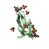
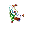
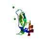
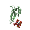
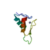
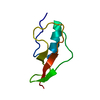
- PDB-2yw8: Crystal structure of human RUN and FYVE domain-containing protein -

+
Open data


ID or keywords:

Loading...

-
Basic information

Entry
Database: PDB / ID: 2yw8
TitleCrystal structure of human RUN and FYVE domain-containing protein
ComponentsRUN and FYVE domain-containing protein 1
KeywordsMETAL BINDING PROTEIN / STRUCTURE GENOMICS / FYVE domain / Structural Genomics / NPPSFA / National Project on Protein Structural and Functional Analyses / RIKEN Structural Genomics/Proteomics Initiative / RSGI
Function / homology
Function and homology information


small GTPase-mediated signal transduction / Synthesis of PIPs at the plasma membrane / regulation of endocytosis / SH2 domain binding / SH3 domain binding / endocytosis / protein transport / early endosome membrane / endosome / nuclear speck ...small GTPase-mediated signal transduction / Synthesis of PIPs at the plasma membrane / regulation of endocytosis / SH2 domain binding / SH3 domain binding / endocytosis / protein transport / early endosome membrane / endosome / nuclear speck / intracellular membrane-bounded organelle / lipid binding / identical protein binding / metal ion binding / cytosol / cytoplasm
Similarity search - Function
: / : / : / RUN / RUN domain / RUN domain superfamily / RUN domain / RUN domain profile. / FYVE zinc finger / FYVE zinc finger ...: / : / : / RUN / RUN domain / RUN domain superfamily / RUN domain / RUN domain profile. / FYVE zinc finger / FYVE zinc finger / Protein present in Fab1, YOTB, Vac1, and EEA1 / Zinc finger, FYVE-related / Zinc finger FYVE/FYVE-related type profile. / Zinc/RING finger domain, C3HC4 (zinc finger) / Herpes Virus-1 / Zinc finger, RING-type / Zinc finger, FYVE/PHD-type / Zinc finger, RING/FYVE/PHD-type / 2-Layer Sandwich / Alpha Beta
Similarity search - Domain/homology
RUN and FYVE domain-containing protein 1
Similarity search - Component
Biological speciesHomo sapiens (human)
MethodX-RAY DIFFRACTION / SYNCHROTRON / MOLECULAR REPLACEMENT / Resolution: 3 Å
AuthorsWang, H. / Kishishita, S. / Murayama, K. / Takemoto, C. / Terada, T. / Shirouzu, M. / RIKEN Structural Genomics/Proteomics Initiative (RSGI)
CitationJournal: To be Published
Title: Crystal structure of human RUN and FYVE domain-containing protein
Authors: Wang, H. / Kishishita, S. / Murayama, T. / Takemoto, C. / Terada, T. / Chen, L. / Fu, Z.Q. / Chrzas, J. / Wang, B.C. / Shirouzu, M. / Yokoyama, S.
History
DepositionApr 20, 2007Deposition site: PDBJ / Processing site: PDBJ
Revision 1.0Apr 22, 2008Provider: repository / Type: Initial release
Revision 1.1Jul 13, 2011Group: Version format compliance
Revision 1.2Oct 25, 2023Group: Data collection / Database references ...Data collection / Database references / Derived calculations / Refinement description
Category: chem_comp_atom / chem_comp_bond ...chem_comp_atom / chem_comp_bond / database_2 / pdbx_initial_refinement_model / pdbx_struct_conn_angle / struct_conn / struct_site
Item: _database_2.pdbx_DOI / _database_2.pdbx_database_accession ..._database_2.pdbx_DOI / _database_2.pdbx_database_accession / _pdbx_struct_conn_angle.ptnr1_auth_seq_id / _pdbx_struct_conn_angle.ptnr1_label_seq_id / _pdbx_struct_conn_angle.ptnr2_auth_seq_id / _pdbx_struct_conn_angle.ptnr2_label_asym_id / _pdbx_struct_conn_angle.ptnr3_auth_seq_id / _pdbx_struct_conn_angle.ptnr3_label_seq_id / _pdbx_struct_conn_angle.value / _struct_conn.pdbx_dist_value / _struct_conn.ptnr1_auth_comp_id / _struct_conn.ptnr1_auth_seq_id / _struct_conn.ptnr1_label_asym_id / _struct_conn.ptnr1_label_atom_id / _struct_conn.ptnr1_label_comp_id / _struct_conn.ptnr1_label_seq_id / _struct_conn.ptnr2_auth_comp_id / _struct_conn.ptnr2_auth_seq_id / _struct_conn.ptnr2_label_asym_id / _struct_conn.ptnr2_label_atom_id / _struct_conn.ptnr2_label_comp_id / _struct_conn.ptnr2_label_seq_id / _struct_site.pdbx_auth_asym_id / _struct_site.pdbx_auth_comp_id / _struct_site.pdbx_auth_seq_id

-
Structure visualization

Structure viewerMolecule:
MolmilJmol/JSmol

Downloads & links

-
Assembly

Deposited unit
A: RUN and FYVE domain-containing protein 1
hetero molecules


Theoretical massNumber of molelcules
Total (without water)9,5744
Polymers9,3471
Non-polymers2273
Water61334
1


  • Idetical with deposited unit
  • defined by author&software
TypeNameSymmetry operationNumber
identity operation1_555x,y,z1
MethodPISA
Unit cell
Length a, b, c (Å)115.150, 115.150, 115.150
Angle α, β, γ (deg.)90.00, 90.00, 90.00
Int Tables number212
Space group name H-MP4332

-
Components

#1: Protein RUN and FYVE domain-containing protein 1 / FYVE-finger protein EIP1 / Zinc finger FYVE domain-containing protein 12 / La-binding protein 1 / ...FYVE-finger protein EIP1 / Zinc finger FYVE domain-containing protein 12 / La-binding protein 1 / Rab4-interacting protein / MS1309 protein


Mass: 9346.705 Da / Num. of mol.: 1 / Fragment: UNP residues 627-708
Source method: isolated from a genetically manipulated source
Source: (gene. exp.) Homo sapiens (human) / Description: cell-free protein synthesis / Gene: RUFY1, RABIP4, ZFYVE12 / Plasmid: PK060327-08 / References: UniProt: Q96T51
#2: Chemical ChemComp-ZN / ZINC ION


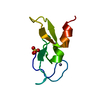
Mass: 65.409 Da / Num. of mol.: 2 / Source method: obtained synthetically / Formula: Zn
#3: Chemical ChemComp-SO4 / SULFATE ION / Sulfate


Mass: 96.063 Da / Num. of mol.: 1 / Source method: obtained synthetically / Formula: SO4
#4: Water ChemComp-HOH / water / Water


Mass: 18.015 Da / Num. of mol.: 34 / Source method: isolated from a natural source / Formula: H2O

-
Experimental details

-
Experiment

ExperimentMethod: X-RAY DIFFRACTION / Number of used crystals: 1

-
Sample preparation

CrystalDensity Matthews: 6.8 Å3/Da / Density % sol: 81.92 %
Crystal growTemperature: 293 K / Method: vapor diffusion, hanging drop / pH: 6.5
Details: Bis-tris, Ammonium sulfate, pH6.5, VAPOR DIFFUSION, HANGING DROP, temperature 293K

-
Data collection

DiffractionMean temperature: 100 K
Diffraction sourceSource: SYNCHROTRON / Site: APS / Beamline: 22-ID / Wavelength: 1.2752 Å
DetectorType: MARMOSAIC 300 mm CCD / Detector: CCD / Date: Feb 5, 2007
RadiationProtocol: SINGLE WAVELENGTH / Monochromatic (M) / Laue (L): M / Scattering type: x-ray
Radiation wavelengthWavelength: 1.2752 Å / Relative weight: 1
ReflectionResolution: 3→50 Å / Num. obs: 5650 / % possible obs: 100 % / Observed criterion σ(I): -3 / Redundancy: 40.9 % / Rsym value: 0.153 / Net I/σ(I): 30.2
Reflection shellResolution: 3→3.11 Å / Redundancy: 42.5 % / Mean I/σ(I) obs: 12.5 / Num. unique all: 544 / Rsym value: 0.414 / % possible all: 100

-
Processing

Software
NameVersionClassification
CNS1.1refinement
HKL-2000data collection
HKL-2000data reduction
HKL-2000data scaling
MOLREPphasing
RefinementMethod to determine structure: MOLECULAR REPLACEMENT
Starting model: PDB ENTRY 1VFY
Resolution: 3→47.01 Å / Rfactor Rfree error: 0.013 / Data cutoff high absF: 44214.21 / Data cutoff low absF: 0 / Isotropic thermal model: RESTRAINED / Cross valid method: THROUGHOUT / σ(F): 0
RfactorNum. reflection% reflectionSelection details
Rfree0.224 314 5.6 %RANDOM
Rwork0.198 ---
obs0.198 5593 99.3 %-
Solvent computationSolvent model: FLAT MODEL / Bsol: 31.5452 Å2 / ksol: 0.356852 e/Å3
Displacement parametersBiso mean: 50.1 Å2
Baniso -1Baniso -2Baniso -3
1-0 Å20 Å20 Å2
2--0 Å20 Å2
3---0 Å2
Refine analyze
FreeObs
Luzzati coordinate error0.35 Å0.3 Å
Luzzati d res low-5 Å
Luzzati sigma a0.33 Å0.33 Å
Refinement stepCycle: LAST / Resolution: 3→47.01 Å
ProteinNucleic acidLigandSolventTotal
Num. atoms487 0 7 34 528
Refine LS restraints
Refine-IDTypeDev ideal
X-RAY DIFFRACTIONc_bond_d0.011
X-RAY DIFFRACTIONc_angle_deg1.8
X-RAY DIFFRACTIONc_dihedral_angle_d25.1
X-RAY DIFFRACTIONc_improper_angle_d0.97
LS refinement shellResolution: 3→3.19 Å / Rfactor Rfree error: 0.042 / Total num. of bins used: 6
RfactorNum. reflection% reflection
Rfree0.308 53 6 %
Rwork0.26 832 -
obs--97.6 %
Xplor file
Refine-IDSerial noParam fileTopol file
X-RAY DIFFRACTION1protein_rep.paramprotein.top
X-RAY DIFFRACTION2water_rep.param
X-RAY DIFFRACTION3ion.param

+
About Yorodumi

-
News

-
Feb 9, 2022. New format data for meta-information of EMDB entries

New format data for meta-information of EMDB entries

  • Version 3 of the EMDB header file is now the official format.
  • The previous official version 1.9 will be removed from the archive.

Related info.:EMDB header

External links:wwPDB to switch to version 3 of the EMDB data model

-
Aug 12, 2020. Covid-19 info

Covid-19 info

URL: https://pdbj.org/emnavi/covid19.php

New page: Covid-19 featured information page in EM Navigator.

Related info.:Covid-19 info / Mar 5, 2020. Novel coronavirus structure data

+
Mar 5, 2020. Novel coronavirus structure data

Novel coronavirus structure data

Related info.:Yorodumi Speices / Aug 12, 2020. Covid-19 info

External links:COVID-19 featured content - PDBj / Molecule of the Month (242):Coronavirus Proteases

+
Jan 31, 2019. EMDB accession codes are about to change! (news from PDBe EMDB page)

EMDB accession codes are about to change! (news from PDBe EMDB page)

  • The allocation of 4 digits for EMDB accession codes will soon come to an end. Whilst these codes will remain in use, new EMDB accession codes will include an additional digit and will expand incrementally as the available range of codes is exhausted. The current 4-digit format prefixed with “EMD-” (i.e. EMD-XXXX) will advance to a 5-digit format (i.e. EMD-XXXXX), and so on. It is currently estimated that the 4-digit codes will be depleted around Spring 2019, at which point the 5-digit format will come into force.
  • The EM Navigator/Yorodumi systems omit the EMD- prefix.

Related info.:Q: What is EMD? / ID/Accession-code notation in Yorodumi/EM Navigator

External links:EMDB Accession Codes are Changing Soon! / Contact to PDBj

+
Jul 12, 2017. Major update of PDB

Major update of PDB

  • wwPDB released updated PDB data conforming to the new PDBx/mmCIF dictionary.
  • This is a major update changing the version number from 4 to 5, and with Remediation, in which all the entries are updated.
  • In this update, many items about electron microscopy experimental information are reorganized (e.g. em_software).
  • Now, EM Navigator and Yorodumi are based on the updated data.

External links:wwPDB Remediation / Enriched Model Files Conforming to OneDep Data Standards Now Available in the PDB FTP Archive

-
Yorodumi

Thousand views of thousand structures

  • Yorodumi is a browser for structure data from EMDB, PDB, SASBDB, etc.
  • This page is also the successor to EM Navigator detail page, and also detail information page/front-end page for Omokage search.
  • The word "yorodu" (or yorozu) is an old Japanese word meaning "ten thousand". "mi" (miru) is to see.

Related info.:EMDB / PDB / SASBDB / Comparison of 3 databanks / Yorodumi Search / Aug 31, 2016. New EM Navigator & Yorodumi / Yorodumi Papers / Jmol/JSmol / Function and homology information / Changes in new EM Navigator and Yorodumi

Read more