 Movie
Movie Controller


+ Open data

- Basic information
| Entry | Database: PDB / ID: 2yhj | ||||||
|---|---|---|---|---|---|---|---|
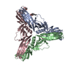
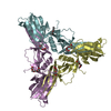
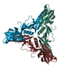
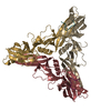
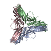
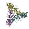
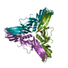
| Title | Clostridium perfringens Enterotoxin at 4.0 Angstrom Resolution | ||||||
 Components Components | HEAT-LABILE ENTEROTOXIN B CHAIN | ||||||
 Keywords Keywords | TOXIN / PORE-FORMING TOXIN / FOOD-POISONING / ANTIBIOTIC-ASSOCIATED DIARRHOEA | ||||||
| Function / homology |  Function and homology information Function and homology information | ||||||
| Biological species |   CLOSTRIDIUM PERFRINGENS (bacteria) CLOSTRIDIUM PERFRINGENS (bacteria) | ||||||
| Method |  X-RAY DIFFRACTION / SYNCHROTRON / MOLECULAR REPLACEMENT / Resolution: 4 Å X-RAY DIFFRACTION / SYNCHROTRON / MOLECULAR REPLACEMENT / Resolution: 4 Å | ||||||
 Authors Authors | Briggs, D.C. / Naylor, C.E. / Smedley III, J.G. / McClane, B.A. / Basak, A.K. | ||||||
 Citation Citation | Journal: J.Mol.Biol. / Year: 2011 Title: Structure of the Food-Poisoning Clostridium Perfringens Enterotoxin Reveals Similarity to the Aerolysin-Like Pore-Forming Toxins Authors: Briggs, D.C. / Naylor, C.E. / Smedley III, J.G. / Lukoyanova, N. / Robertson, S. / Mcclane, B.A. / Basak, A.K. #1: Journal: Acta Crystallogr.,Sect.F / Year: 2010 Title: Crystallization and Preliminary Crystallographic Analysis of the Clostridium Perfringens Enterotoxin. Authors: Briggs, D.C. / Smedley III, J.G. / Mcclane, B.A. / Basak, A.K. | ||||||
| History |
|
- Structure visualization
| Structure viewer | Molecule: MolmilJmol/JSmol |
|---|
- Downloads & links
-Download
| PDBx/mmCIF format | 2yhj.cif.gz | 121.3 KB | Display | PDBx/mmCIF format |
|---|---|---|---|---|
| PDB format | pdb2yhj.ent.gz | 95.1 KB | Display | PDB format |
| PDBx/mmJSON format | 2yhj.json.gz | Tree view | PDBx/mmJSON format | |
| Others |  Other downloads Other downloads |
-Validation report
| Arichive directory | https://data.pdbj.org/pub/pdb/validation_reports/yh/2yhjftp://data.pdbj.org/pub/pdb/validation_reports/yh/2yhj | HTTPS FTP |
|---|
-Related structure data
| Related structure data |  2xh6SC S: Starting model for refinement C: citing same article ( |
|---|---|
| Similar structure data |

-Links
 PDBj
PDBj- Assembly
| Deposited unit | 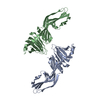
| ||||||||
|---|---|---|---|---|---|---|---|---|---|
| 1 | 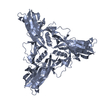
| ||||||||
| 2 | 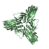
| ||||||||
| Unit cell |
| ||||||||
| Noncrystallographic symmetry (NCS) | NCS oper: (Code: given Matrix: (-0.00405, -0.99991, 0.01284), Vector: |
-Components
| #1: Protein | Mass: 35345.207 Da / Num. of mol.: 2 / Source method: isolated from a natural source / Source: (natural) CLOSTRIDIUM PERFRINGENS (bacteria) / Strain: NCTC 8239 / References: UniProt: P01558 |
|---|
-Experimental details
-Experiment
| Experiment | Method: X-RAY DIFFRACTION / Number of used crystals: 1 |
|---|
- Sample preparation
| Crystal | Density Matthews: 4.8 Å3/Da / Density % sol: 75 % / Description: NONE |
|---|---|
| Crystal grow | pH: 4.3 Details: 10 MG/ML PROTEIN IN WATER EQUILIBRATED AGAINST 2.0 M HEXANE-1,2-DIOL AND 10 MM ZNCL2 IN 50 MM CITRATE, PH 4.3 . |
-Data collection
| Diffraction | Mean temperature: 100 K |
|---|---|
| Diffraction source | Source: SYNCHROTRON / Site: ESRF  / Beamline: ID14-4 / Wavelength: 1.2823 / Beamline: ID14-4 / Wavelength: 1.2823 |
| Detector | Type: ADSC CCD / Detector: CCD / Date: Jul 2, 2003 / Details: MIRRORS |
| Radiation | Monochromator: SI CRYSTAL / Protocol: SINGLE WAVELENGTH / Monochromatic (M) / Laue (L): M / Scattering type: x-ray |
| Radiation wavelength | Wavelength: 1.2823 Å / Relative weight: 1 |
| Reflection | Resolution: 4→70 Å / Num. obs: 11712 / % possible obs: 99.9 % / Observed criterion σ(I): -3 / Redundancy: 10 % / Biso Wilson estimate: 85.23 Å2 / Rmerge(I) obs: 0.17 / Net I/σ(I): 16.7 |
| Reflection shell | Resolution: 4→4.2 Å / Redundancy: 7.4 % / Rmerge(I) obs: 0.62 / Mean I/σ(I) obs: 3.2 / % possible all: 99.5 |
- Processing
| Software |
| ||||||||||||||||||||||||||||||||||||||||||||||||||||||||||||||||||||||||||||||||||||||||||||||||||||||||||||||||||
|---|---|---|---|---|---|---|---|---|---|---|---|---|---|---|---|---|---|---|---|---|---|---|---|---|---|---|---|---|---|---|---|---|---|---|---|---|---|---|---|---|---|---|---|---|---|---|---|---|---|---|---|---|---|---|---|---|---|---|---|---|---|---|---|---|---|---|---|---|---|---|---|---|---|---|---|---|---|---|---|---|---|---|---|---|---|---|---|---|---|---|---|---|---|---|---|---|---|---|---|---|---|---|---|---|---|---|---|---|---|---|---|---|---|---|---|
| Refinement | Method to determine structure: MOLECULAR REPLACEMENT Starting model: PDB ENTRY 2XH6, CHAIN A Resolution: 4→65.2 Å / Cor.coef. Fo:Fc: 0.8563 / Cor.coef. Fo:Fc free: 0.7484 / Cross valid method: THROUGHOUT / σ(F): 0 / Details: THERE IS NO ELECTRON DENSITY FOR RESIDUES 1-34
| ||||||||||||||||||||||||||||||||||||||||||||||||||||||||||||||||||||||||||||||||||||||||||||||||||||||||||||||||||
| Displacement parameters | Biso mean: 115.4 Å2
| ||||||||||||||||||||||||||||||||||||||||||||||||||||||||||||||||||||||||||||||||||||||||||||||||||||||||||||||||||
| Refine analyze | Luzzati coordinate error obs: 1.025 Å | ||||||||||||||||||||||||||||||||||||||||||||||||||||||||||||||||||||||||||||||||||||||||||||||||||||||||||||||||||
| Refinement step | Cycle: LAST / Resolution: 4→65.2 Å
| ||||||||||||||||||||||||||||||||||||||||||||||||||||||||||||||||||||||||||||||||||||||||||||||||||||||||||||||||||
| Refine LS restraints |
| ||||||||||||||||||||||||||||||||||||||||||||||||||||||||||||||||||||||||||||||||||||||||||||||||||||||||||||||||||
| LS refinement shell | Resolution: 4→4.38 Å / Total num. of bins used: 6
|
