 Movie
Movie Controller

[English] 日本語
 Yorodumi
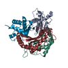
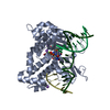
- PDB-2yf2: Crystal structure of the oligomerisation domain of C4b-binding pr... -
+ Open data

- Basic information
| Entry | Database: PDB / ID: 2yf2 | ||||||
|---|---|---|---|---|---|---|---|
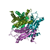
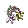
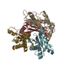
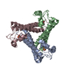
| Title | Crystal structure of the oligomerisation domain of C4b-binding protein from Gallus gallus | ||||||
 Components Components | C4B BINDING PROTEIN | ||||||
 Keywords Keywords | IMMUNE SYSTEM / COMPLEMENT SYSTEM | ||||||
| Function / homology | Single alpha-helices involved in coiled-coils or other helix-helix interfaces - #3730 / Single alpha-helices involved in coiled-coils or other helix-helix interfaces / Up-down Bundle / Mainly Alpha / ACETATE ION / :  Function and homology information Function and homology information | ||||||
| Biological species |  | ||||||
| Method |  X-RAY DIFFRACTION / SYNCHROTRON / SIRAS / Resolution: 2.24 Å X-RAY DIFFRACTION / SYNCHROTRON / SIRAS / Resolution: 2.24 Å | ||||||
 Authors Authors | Caesar, J.J.E. / Hill, F. / Lea, S.M. | ||||||
 Citation Citation | Journal: To be Published Title: Crystal Structure of the Oligomerisation Domain of C4B-Binding Protein from Gallus Gallus Authors: Caesar, J.J.E. / Hill, F. / Lea, S.M. | ||||||
| History |
|
- Structure visualization
| Structure viewer | Molecule: MolmilJmol/JSmol |
|---|
- Downloads & links
-Download
| PDBx/mmCIF format | 2yf2.cif.gz | 87.7 KB | Display | PDBx/mmCIF format |
|---|---|---|---|---|
| PDB format | pdb2yf2.ent.gz | 68.8 KB | Display | PDB format |
| PDBx/mmJSON format | 2yf2.json.gz | Tree view | PDBx/mmJSON format | |
| Others |  Other downloads Other downloads |
-Validation report
| Arichive directory | https://data.pdbj.org/pub/pdb/validation_reports/yf/2yf2ftp://data.pdbj.org/pub/pdb/validation_reports/yf/2yf2 | HTTPS FTP |
|---|
-Related structure data
| Similar structure data |
|---|


-Links
 PDBj
PDBj- Assembly
| Deposited unit | 
| ||||||||||||||||||||||||||||
|---|---|---|---|---|---|---|---|---|---|---|---|---|---|---|---|---|---|---|---|---|---|---|---|---|---|---|---|---|---|
| 1 |
| ||||||||||||||||||||||||||||
| Unit cell |
| ||||||||||||||||||||||||||||
| Components on special symmetry positions |
| ||||||||||||||||||||||||||||
| Noncrystallographic symmetry (NCS) | NCS oper:
|
-Components
| #1: Protein | Mass: 7304.416 Da / Num. of mol.: 7 / Fragment: OLIGOMERISATION DOMAIN, RESIDUES 395-457 / Mutation: YES Source method: isolated from a genetically manipulated source Source: (gene. exp.)  #2: Chemical | ChemComp-EDO /   Mass: 62.068 Da / Num. of mol.: 19 / Source method: obtained synthetically / Formula: C2H6O2 Mass: 62.068 Da / Num. of mol.: 19 / Source method: obtained synthetically / Formula: C2H6O2#3: Chemical | ChemComp-ACT /   Mass: 59.044 Da / Num. of mol.: 11 / Source method: obtained synthetically / Formula: C2H3O2 Mass: 59.044 Da / Num. of mol.: 11 / Source method: obtained synthetically / Formula: C2H3O2#4: Water | ChemComp-HOH / |  Mass: 18.015 Da / Num. of mol.: 71 / Source method: isolated from a natural source / Formula: H2O Mass: 18.015 Da / Num. of mol.: 71 / Source method: isolated from a natural source / Formula: H2OCompound details | ENGINEERED RESIDUE IN CHAIN A, CYS 395 TO SER ENGINEERED RESIDUE IN CHAIN B, CYS 395 TO SER ...ENGINEERED | Has protein modification | Y | |
|---|
-Experimental details
-Experiment
| Experiment | Method: X-RAY DIFFRACTION / Number of used crystals: 1 |
|---|
- Sample preparation
| Crystal | Density Matthews: 3.07 Å3/Da / Density % sol: 59.9 % / Description: NONE |
|---|---|
| Crystal grow | Details: 0.2M AMMONIUM ACETATE, 0.1M SODIUM ACETATE, 8% PEG 3350, PH 4.5 |
-Data collection
| Diffraction | Mean temperature: 120 K |
|---|---|
| Diffraction source | Source: SYNCHROTRON / Site: Diamond  / Beamline: I03 / Wavelength: 0.9793 / Beamline: I03 / Wavelength: 0.9793 |
| Detector | Type: ADSC CCD / Detector: CCD / Date: Apr 24, 2010 |
| Radiation | Protocol: SINGLE WAVELENGTH / Monochromatic (M) / Laue (L): M / Scattering type: x-ray |
| Radiation wavelength | Wavelength: 0.9793 Å / Relative weight: 1 |
| Reflection | Resolution: 2.24→85.46 Å / Num. obs: 23669 / % possible obs: 96 % / Observed criterion σ(I): 2 / Redundancy: 4.3 % / Biso Wilson estimate: 38.19 Å2 / Rmerge(I) obs: 0.06 / Net I/σ(I): 13.5 |
| Reflection shell | Resolution: 2.24→2.36 Å / Redundancy: 2.8 % / Rmerge(I) obs: 0.49 / Mean I/σ(I) obs: 2.2 / % possible all: 79.6 |
- Processing
| Software |
| ||||||||||||||||||||||||||||||||||||||||||||||||||||||||||||||||||||||||||||||||||||||||||||||||||||||||||||||||||
|---|---|---|---|---|---|---|---|---|---|---|---|---|---|---|---|---|---|---|---|---|---|---|---|---|---|---|---|---|---|---|---|---|---|---|---|---|---|---|---|---|---|---|---|---|---|---|---|---|---|---|---|---|---|---|---|---|---|---|---|---|---|---|---|---|---|---|---|---|---|---|---|---|---|---|---|---|---|---|---|---|---|---|---|---|---|---|---|---|---|---|---|---|---|---|---|---|---|---|---|---|---|---|---|---|---|---|---|---|---|---|---|---|---|---|---|
| Refinement | Method to determine structure: SIRAS Starting model: NONE Resolution: 2.24→55.06 Å / Cor.coef. Fo:Fc: 0.9068 / Cor.coef. Fo:Fc free: 0.8888 / SU R Cruickshank DPI: 0.261 / Cross valid method: THROUGHOUT / σ(F): 0 / SU R Blow DPI: 0.259 / SU Rfree Blow DPI: 0.191 / SU Rfree Cruickshank DPI: 0.193 Details: IDEAL-DIST CONTACT TERM CONTACT SETUP. ALL ATOMS HAVE CCP4 ATOM TYPE FROM LIBRARY.
| ||||||||||||||||||||||||||||||||||||||||||||||||||||||||||||||||||||||||||||||||||||||||||||||||||||||||||||||||||
| Displacement parameters | Biso mean: 53.53 Å2
| ||||||||||||||||||||||||||||||||||||||||||||||||||||||||||||||||||||||||||||||||||||||||||||||||||||||||||||||||||
| Refinement step | Cycle: LAST / Resolution: 2.24→55.06 Å
| ||||||||||||||||||||||||||||||||||||||||||||||||||||||||||||||||||||||||||||||||||||||||||||||||||||||||||||||||||
| Refine LS restraints |
| ||||||||||||||||||||||||||||||||||||||||||||||||||||||||||||||||||||||||||||||||||||||||||||||||||||||||||||||||||
| LS refinement shell | Resolution: 2.24→2.34 Å / Total num. of bins used: 12
|
