 Movie
Movie Controller

[English] 日本語
 Yorodumi
- PDB-2ycl: complete structure of the corrinoid,iron-sulfur protein including... -
+ Open data

- Basic information
| Entry | Database: PDB / ID: 2ycl | ||||||
|---|---|---|---|---|---|---|---|
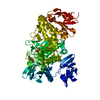
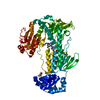
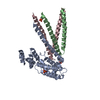
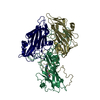
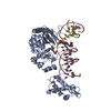
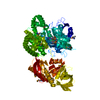
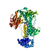
| Title | complete structure of the corrinoid,iron-sulfur protein including the N-terminal domain with a 4Fe-4S cluster | ||||||
 Components Components |
| ||||||
 Keywords Keywords | TRANSFERASE | ||||||
| Function / homology |  Function and homology information Function and homology informationacetyl-CoA catabolic process / methyltransferase activity / 4 iron, 4 sulfur cluster binding / iron ion binding Similarity search - Function | ||||||
| Biological species |   CARBOXYDOTHERMUS HYDROGENOFORMANS (bacteria) CARBOXYDOTHERMUS HYDROGENOFORMANS (bacteria) | ||||||
| Method |  X-RAY DIFFRACTION / SYNCHROTRON / MOLECULAR REPLACEMENT / Resolution: 1.95 Å X-RAY DIFFRACTION / SYNCHROTRON / MOLECULAR REPLACEMENT / Resolution: 1.95 Å | ||||||
 Authors Authors | Goetzl, S. / Jeoung, J.H. / Hennig, S.E. / Dobbek, H. | ||||||
 Citation Citation | Journal: J.Mol.Biol. / Year: 2011 Title: Structural Basis for Electron and Methyl-Group Transfer in a Methyltransferase System Operating in the Reductive Acetyl-Coa Pathway Authors: Goetzl, S. / Jeoung, J.H. / Hennig, S.E. / Dobbek, H. | ||||||
| History |
|
- Structure visualization
| Structure viewer | Molecule: MolmilJmol/JSmol |
|---|
- Downloads & links
-Download
| PDBx/mmCIF format | 2ycl.cif.gz | 181 KB | Display | PDBx/mmCIF format |
|---|---|---|---|---|
| PDB format | pdb2ycl.ent.gz | 141.1 KB | Display | PDB format |
| PDBx/mmJSON format | 2ycl.json.gz | Tree view | PDBx/mmJSON format | |
| Others |  Other downloads Other downloads |
-Validation report
| Arichive directory | https://data.pdbj.org/pub/pdb/validation_reports/yc/2yclftp://data.pdbj.org/pub/pdb/validation_reports/yc/2ycl | HTTPS FTP |
|---|
-Related structure data
| Related structure data |  2yciC  2ycjC  2yckC 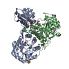 2h9aS C: citing same article ( S: Starting model for refinement |
|---|---|
| Similar structure data |


-Links
 PDBj
PDBj- Assembly
| Deposited unit | 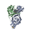
| ||||||||
|---|---|---|---|---|---|---|---|---|---|
| 1 |
| ||||||||
| Unit cell |
| ||||||||
| Components on special symmetry positions |
|
-Components
| #1: Protein | Mass: 48469.820 Da / Num. of mol.: 1 Source method: isolated from a genetically manipulated source Source: (gene. exp.) CARBOXYDOTHERMUS HYDROGENOFORMANS (bacteria)Strain: Z-2901 / Production host: |
|---|---|
| #2: Protein | Mass: 33910.355 Da / Num. of mol.: 1 Source method: isolated from a genetically manipulated source Source: (gene. exp.) CARBOXYDOTHERMUS HYDROGENOFORMANS (bacteria)Strain: Z-2901 / Production host: |
| #3: Chemical | ChemComp-B12 /   Mass: 1330.356 Da / Num. of mol.: 1 / Source method: obtained synthetically / Formula: C62H89CoN13O14P Mass: 1330.356 Da / Num. of mol.: 1 / Source method: obtained synthetically / Formula: C62H89CoN13O14P |
| #4: Chemical | ChemComp-SF4 /   Mass: 351.640 Da / Num. of mol.: 1 / Source method: obtained synthetically / Formula: Fe4S4 Mass: 351.640 Da / Num. of mol.: 1 / Source method: obtained synthetically / Formula: Fe4S4 |
| #5: Water | ChemComp-HOH /  Mass: 18.015 Da / Num. of mol.: 795 / Source method: isolated from a natural source / Formula: H2O Mass: 18.015 Da / Num. of mol.: 795 / Source method: isolated from a natural source / Formula: H2O |
-Experimental details
-Experiment
| Experiment | Method: X-RAY DIFFRACTION |
|---|
- Sample preparation
| Crystal | Density Matthews: 2.02 Å3/Da / Density % sol: 39 % / Description: NONE |
|---|
-Data collection
| Diffraction | Mean temperature: 100 K |
|---|---|
| Diffraction source | Source: SYNCHROTRON / Site: BESSY  / Beamline: 14.2 / Wavelength: 0.9184 / Beamline: 14.2 / Wavelength: 0.9184 |
| Detector | Type: MARRESEARCH / Detector: CCD |
| Radiation | Protocol: SINGLE WAVELENGTH / Monochromatic (M) / Laue (L): M / Scattering type: x-ray |
| Radiation wavelength | Wavelength: 0.9184 Å / Relative weight: 1 |
| Reflection | Resolution: 1.95→35 Å / Num. obs: 48286 / % possible obs: 99.3 % / Observed criterion σ(I): 2 / Redundancy: 3.9 % / Biso Wilson estimate: 18.68 Å2 / Rmerge(I) obs: 0.1 / Net I/σ(I): 10.36 |
| Reflection shell | Resolution: 1.95→2 Å / Redundancy: 3.8 % / Rmerge(I) obs: 0.53 / Mean I/σ(I) obs: 2.55 / % possible all: 99.4 |
- Processing
| Software |
| ||||||||||||||||||||||||||||||||||||||||||||||||||||||||||||||||||||||||||||||||||||||||||||||||||||||||||||||||||||||||||||||
|---|---|---|---|---|---|---|---|---|---|---|---|---|---|---|---|---|---|---|---|---|---|---|---|---|---|---|---|---|---|---|---|---|---|---|---|---|---|---|---|---|---|---|---|---|---|---|---|---|---|---|---|---|---|---|---|---|---|---|---|---|---|---|---|---|---|---|---|---|---|---|---|---|---|---|---|---|---|---|---|---|---|---|---|---|---|---|---|---|---|---|---|---|---|---|---|---|---|---|---|---|---|---|---|---|---|---|---|---|---|---|---|---|---|---|---|---|---|---|---|---|---|---|---|---|---|---|---|
| Refinement | Method to determine structure: MOLECULAR REPLACEMENT Starting model: PDB ENTRY 2H9A Resolution: 1.95→33.745 Å / SU ML: 0.27 / σ(F): 1.99 / Phase error: 27.63 / Stereochemistry target values: ML
| ||||||||||||||||||||||||||||||||||||||||||||||||||||||||||||||||||||||||||||||||||||||||||||||||||||||||||||||||||||||||||||||
| Solvent computation | Shrinkage radii: 0.65 Å / VDW probe radii: 0.8 Å / Solvent model: FLAT BULK SOLVENT MODEL / Bsol: 50.296 Å2 / ksol: 0.427 e/Å3 | ||||||||||||||||||||||||||||||||||||||||||||||||||||||||||||||||||||||||||||||||||||||||||||||||||||||||||||||||||||||||||||||
| Displacement parameters |
| ||||||||||||||||||||||||||||||||||||||||||||||||||||||||||||||||||||||||||||||||||||||||||||||||||||||||||||||||||||||||||||||
| Refinement step | Cycle: LAST / Resolution: 1.95→33.745 Å
| ||||||||||||||||||||||||||||||||||||||||||||||||||||||||||||||||||||||||||||||||||||||||||||||||||||||||||||||||||||||||||||||
| Refine LS restraints |
| ||||||||||||||||||||||||||||||||||||||||||||||||||||||||||||||||||||||||||||||||||||||||||||||||||||||||||||||||||||||||||||||
| LS refinement shell |
|
