Movie
Movie Controller

[English] 日本語
 Yorodumi
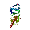
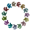
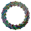
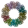
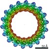
- PDB-2y9k: Three-dimensional model of Salmonella's needle complex at subnano... -
+ Open data

- Basic information
| Entry | Database: PDB / ID: 2y9k | ||||||
|---|---|---|---|---|---|---|---|
| Title | Three-dimensional model of Salmonella's needle complex at subnanometer resolution | ||||||
 Components Components | PROTEIN INVG | ||||||
 Keywords Keywords | PROTEIN TRANSPORT / TYPE III SECRETION SYSTEM / OUTER MEMBRANE RING / SECRETIN FAMILY / C15 FOLD | ||||||
| Function / homology |  Function and homology information Function and homology informationtype III protein secretion system complex / type II protein secretion system complex / protein secretion by the type III secretion system / protein secretion / cell outer membrane / identical protein binding Similarity search - Function | ||||||
| Biological species |  SALMONELLA ENTERICA SUBSP. ENTERICA SEROVAR TYPHIMURIUM (bacteria) SALMONELLA ENTERICA SUBSP. ENTERICA SEROVAR TYPHIMURIUM (bacteria) | ||||||
| Method | ELECTRON MICROSCOPY / single particle reconstruction / cryo EM / Resolution: 8.3 Å | ||||||
 Authors Authors | Schraidt, O. / Marlovits, T.C. | ||||||
 Citation Citation | Journal: Science / Year: 2011 Title: Three-dimensional model of Salmonella's needle complex at subnanometer resolution. Authors: Oliver Schraidt / Thomas C Marlovits /  Abstract: Type III secretion systems (T3SSs) are essential virulence factors used by many Gram-negative bacteria to inject proteins that make eukaryotic host cells accessible to invasion. The T3SS core ...Type III secretion systems (T3SSs) are essential virulence factors used by many Gram-negative bacteria to inject proteins that make eukaryotic host cells accessible to invasion. The T3SS core structure, the needle complex (NC), is a ~3.5 megadalton-sized, oligomeric, membrane-embedded complex. Analyzing cryo-electron microscopy images of top views of NCs or NC substructures from Salmonella typhimurium revealed a 24-fold symmetry for the inner rings and a 15-fold symmetry for the outer rings, giving an overall C3 symmetry. Local refinement and averaging showed the organization of the central core and allowed us to reconstruct a subnanometer composite structure of the NC, which together with confident docking of atomic structures reveal insights into its overall organization and structural requirements during assembly. | ||||||
| History |
|
- Structure visualization
| Movie |
Movie viewer |
|---|---|
| Structure viewer | Molecule: MolmilJmol/JSmol |
- Downloads & links
-Download
| PDBx/mmCIF format | 2y9k.cif.gz | 352.2 KB | Display | PDBx/mmCIF format |
|---|---|---|---|---|
| PDB format | pdb2y9k.ent.gz | 282.6 KB | Display | PDB format |
| PDBx/mmJSON format | 2y9k.json.gz | Tree view | PDBx/mmJSON format | |
| Others |  Other downloads Other downloads |
-Validation report
| Arichive directory | https://data.pdbj.org/pub/pdb/validation_reports/y9/2y9kftp://data.pdbj.org/pub/pdb/validation_reports/y9/2y9k | HTTPS FTP |
|---|
-Related structure data
| Related structure data | 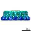 1871MC 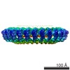 1874C 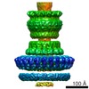 1875C 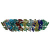 2y9jC C: citing same article ( M: map data used to model this data |
|---|---|
| Similar structure data |
-Links
 PDBj
PDBj
- Assembly
| Deposited unit | 
|
|---|---|
| 1 |
|
-Components
| #1: Protein | Mass: 15616.915 Da / Num. of mol.: 15 / Fragment: N-TERMINAL DOMAIN, RESIDUES 34-170 / Source method: isolated from a natural source Source: (natural) SALMONELLA ENTERICA SUBSP. ENTERICA SEROVAR TYPHIMURIUM (bacteria)References: UniProt: P35672 |
|---|
-Experimental details
-Experiment
| Experiment | Method: ELECTRON MICROSCOPY |
|---|---|
| EM experiment | Aggregation state: PARTICLE / 3D reconstruction method: single particle reconstruction |
- Sample preparation
| Component | Name: NEEDLE COMPLEX / Type: COMPLEX |
|---|---|
| Buffer solution | pH: 7.5 / Details: 10mM Tris-HCl 0.5M NaCl 0.1% LDAO |
| Specimen | Embedding applied: NO / Shadowing applied: NO / Staining applied: NO / Vitrification applied: YES |
| Specimen support | Details: CARBON |
| Vitrification | Cryogen name: ETHANE / Details: LIQUID ETHANE |
- Electron microscopy imaging
| Experimental equipment |  Model: Tecnai Polara / Image courtesy: FEI Company |
|---|---|
| Microscopy | Model: FEI POLARA 300 Details: ACUAL MAGNIFICATION AT CCD 112968, CAMERA PIXEL SIZE 15UM, 1.33 ANGSTROM PER PIXEL, DATA COLLECTED SEMI- AUTOMATICALLY USING POINT-2-POINT (DEVELOPED IN-HOUSE) |
| Electron gun | Electron source:  FIELD EMISSION GUN / Accelerating voltage: 300 kV / Illumination mode: FLOOD BEAM FIELD EMISSION GUN / Accelerating voltage: 300 kV / Illumination mode: FLOOD BEAM |
| Electron lens | Mode: BRIGHT FIELD / Nominal magnification: 93000 X / Nominal defocus max: 2500 nm / Nominal defocus min: 1000 nm / Cs: 2 mm |
| Specimen holder | Tilt angle min: 0 ° |
| Image recording | Film or detector model: GENERIC GATAN (4k x 4k) |
| Radiation wavelength | Relative weight: 1 |
- Processing
| EM software |
| ||||||||||||
|---|---|---|---|---|---|---|---|---|---|---|---|---|---|
| CTF correction | Details: EACH CCD FRAME | ||||||||||||
| Symmetry | Point symmetry: C15 (15 fold cyclic) | ||||||||||||
| 3D reconstruction | Method: PROJECTION MATCHING / Resolution: 8.3 Å / Resolution method: FSC 0.5 CUT-OFF / Actual pixel size: 1.33 Å Details: RESOLUTION 8.3 ANGSTROM (0.5 FSC), 6.7 ANGSTROM (HALF BIT) SUBMISSION BASED ON EXPERIMENTAL DATA FROM EMDB EMD-1871. (DEPOSITION ID: 7820). Symmetry type: POINT | ||||||||||||
| Atomic model building | Protocol: RIGID BODY FIT / Space: REAL / Target criteria: Cross-correlation coefficient / Details: METHOD--RIGID BODY FITTING | ||||||||||||
| Atomic model building | PDB-ID: 3GR5 Accession code: 3GR5 / Source name: PDB / Type: experimental model | ||||||||||||
| Refinement | Highest resolution: 8.3 Å | ||||||||||||
| Refinement step | Cycle: LAST / Highest resolution: 8.3 Å
|