Entry Database : PDB / ID : 2y96Title Structure of human dual-specificity phosphatase 27 DUAL SPECIFICITY PHOSPHATASE DUPD1 Keywords Function / homology Function Domain/homology Component
/ / / / / / / / / / / / / / / / / / / / / / / / / / / / / / / / / / / / / / / / / / / / / / Biological species HOMO SAPIENS (human)Method / / / Resolution : 2.38 Å Authors Lountos, G.T. / Tropea, J.E. / Waugh, D.S. Journal : Acta Crystallogr.,Sect.D / Year : 2011Title : Structure of Human Dual-Specificity Phosphatase at 2.38A ResolutionAuthors : Lountos, G.T. / Tropea, J.E. / Waugh, D.S. History Deposition Feb 11, 2011 Deposition site / Processing site Revision 1.0 Apr 27, 2011 Provider / Type Revision 1.1 May 26, 2011 Group Revision 1.2 Jul 13, 2011 Group Revision 1.3 Dec 20, 2023 Group Data collection / Database references ... Data collection / Database references / Derived calculations / Other / Refinement description Category chem_comp_atom / chem_comp_bond ... chem_comp_atom / chem_comp_bond / database_2 / pdbx_database_status / pdbx_initial_refinement_model / struct_site Item _database_2.pdbx_DOI / _database_2.pdbx_database_accession ... _database_2.pdbx_DOI / _database_2.pdbx_database_accession / _pdbx_database_status.status_code_sf / _struct_site.pdbx_auth_asym_id / _struct_site.pdbx_auth_comp_id / _struct_site.pdbx_auth_seq_id
Show all Show less
 Movie
Movie Controller


 Open data
 Basic information
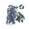
 Components
 Keywords
 Function and homology information
 HOMO SAPIENS (human)
 X-RAY DIFFRACTION /
  Authors
 Citation
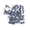
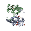
 Structure visualization
 Downloads & links
 Other downloads


 PDBj
PDBj


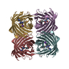
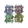
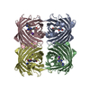
 Assembly


 Mass: 96.063 Da / Num. of mol.: 7 / Source method: obtained synthetically / Formula: SO4
 Mass: 18.015 Da / Num. of mol.: 137 / Source method: isolated from a natural source / Formula: H2O
 Sample preparation
 / Beamline: 22-BM / Wavelength: 1
  Processing