 Movie
Movie Controller

[English] 日本語
 Yorodumi
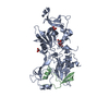
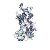
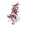
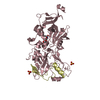
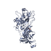
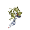
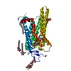
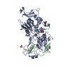
- PDB-2y8r: Crystal structure of apo AMA1 mutant (Tyr230Ala) from Toxoplasma ... -
+ Open data

- Basic information
| Entry | Database: PDB / ID: 2y8r | ||||||
|---|---|---|---|---|---|---|---|
| Title | Crystal structure of apo AMA1 mutant (Tyr230Ala) from Toxoplasma gondii | ||||||
 Components Components | APICAL MEMBRANE ANTIGEN, PUTATIVE | ||||||
 Keywords Keywords | MEMBRANE PROTEIN / MOVING JUNCTION / INVASION | ||||||
| Function / homology |  Function and homology information Function and homology information | ||||||
| Biological species |  | ||||||
| Method |  X-RAY DIFFRACTION / SYNCHROTRON / MOLECULAR REPLACEMENT / Resolution: 2.45 Å X-RAY DIFFRACTION / SYNCHROTRON / MOLECULAR REPLACEMENT / Resolution: 2.45 Å | ||||||
 Authors Authors | Tonkin, M.L. / Roques, M. / Lamarque, M.H. / Pugniere, M. / Douguet, D. / Crawford, J. / Lebrun, M. / Boulanger, M.J. | ||||||
 Citation Citation | Journal: Science / Year: 2011 Title: Host Cell Invasion by Apicomplexan Parasites: Insights from the Co-Structure of Ama1 with a Ron2 Peptide Authors: Tonkin, M.L. / Roques, M. / Lamarque, M.H. / Pugniere, M. / Douguet, D. / Crawford, J. / Lebrun, M. / Boulanger, M.J. | ||||||
| History |
|
- Structure visualization
| Structure viewer | Molecule: MolmilJmol/JSmol |
|---|
- Downloads & links
-Download
| PDBx/mmCIF format | 2y8r.cif.gz | 328.4 KB | Display | PDBx/mmCIF format |
|---|---|---|---|---|
| PDB format | pdb2y8r.ent.gz | 267.1 KB | Display | PDB format |
| PDBx/mmJSON format | 2y8r.json.gz | Tree view | PDBx/mmJSON format | |
| Others |  Other downloads Other downloads |
-Validation report
| Arichive directory | https://data.pdbj.org/pub/pdb/validation_reports/y8/2y8rftp://data.pdbj.org/pub/pdb/validation_reports/y8/2y8r | HTTPS FTP |
|---|
-Related structure data


-Links
 PDBj
PDBj- Assembly
| Deposited unit | 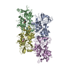
| ||||||||
|---|---|---|---|---|---|---|---|---|---|
| 1 | 
| ||||||||
| 2 | 
| ||||||||
| 3 | 
| ||||||||
| 4 | 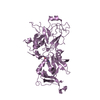
| ||||||||
| Unit cell |
|
-Components
| #1: Protein | Mass: 47921.363 Da / Num. of mol.: 4 / Fragment: DOMAINS I/II/III, RESIDUES 64-484 / Mutation: YES Source method: isolated from a genetically manipulated source Source: (gene. exp.)  TRICHOPLUSIA NI (cabbage looper) / References: UniProt: B9QC59, UniProt: B6KAM0*PLUS TRICHOPLUSIA NI (cabbage looper) / References: UniProt: B9QC59, UniProt: B6KAM0*PLUS#2: Sugar | ChemComp-NAG /   Type: D-saccharide, beta linking / Mass: 221.208 Da / Num. of mol.: 4 Type: D-saccharide, beta linking / Mass: 221.208 Da / Num. of mol.: 4Source method: isolated from a genetically manipulated source Formula: C8H15NO6 #3: Water | ChemComp-HOH / |  Mass: 18.015 Da / Num. of mol.: 529 / Source method: isolated from a natural source / Formula: H2O Mass: 18.015 Da / Num. of mol.: 529 / Source method: isolated from a natural source / Formula: H2OCompound details | ENGINEERED RESIDUE IN CHAIN A, TYR 230 TO ALA ENGINEERED RESIDUE IN CHAIN B, TYR 230 TO ALA ...ENGINEERED | Has protein modification | Y | |
|---|
-Experimental details
-Experiment
| Experiment | Method: X-RAY DIFFRACTION |
|---|
- Sample preparation
| Crystal | Density Matthews: 2.12 Å3/Da / Density % sol: 41.89 % / Description: NONE |
|---|---|
| Crystal grow | Details: 21% PEG3350, 100 MM TRIS PH 7.4 |
-Data collection
| Diffraction | Mean temperature: 100 K |
|---|---|
| Diffraction source | Source: SYNCHROTRON / Site: SSRL  / Beamline: BL9-2 / Wavelength: 0.9795 / Beamline: BL9-2 / Wavelength: 0.9795 |
| Detector | Type: MARMOSAIC 325 mm CCD / Detector: CCD |
| Radiation | Protocol: SINGLE WAVELENGTH / Monochromatic (M) / Laue (L): M / Scattering type: x-ray |
| Radiation wavelength | Wavelength: 0.9795 Å / Relative weight: 1 |
| Reflection | Resolution: 2.35→38.59 Å / Num. obs: 63415 / % possible obs: 96.9 % / Observed criterion σ(I): 2 / Redundancy: 2.4 % / Rmerge(I) obs: 0.1 / Net I/σ(I): 7.2 |
| Reflection shell | Resolution: 2.35→2.48 Å / Redundancy: 2.4 % / Rmerge(I) obs: 0.46 / Mean I/σ(I) obs: 1.9 / % possible all: 96.1 |
- Processing
| Software |
| ||||||||||||||||||||||||||||||||||||||||||||||||||||||||||||||||||||||||||||||||||||||||||||||||||||||||||||||||||||||||||||||||||||||||||||||||||||||||||||||||||||||||||||||||||||||
|---|---|---|---|---|---|---|---|---|---|---|---|---|---|---|---|---|---|---|---|---|---|---|---|---|---|---|---|---|---|---|---|---|---|---|---|---|---|---|---|---|---|---|---|---|---|---|---|---|---|---|---|---|---|---|---|---|---|---|---|---|---|---|---|---|---|---|---|---|---|---|---|---|---|---|---|---|---|---|---|---|---|---|---|---|---|---|---|---|---|---|---|---|---|---|---|---|---|---|---|---|---|---|---|---|---|---|---|---|---|---|---|---|---|---|---|---|---|---|---|---|---|---|---|---|---|---|---|---|---|---|---|---|---|---|---|---|---|---|---|---|---|---|---|---|---|---|---|---|---|---|---|---|---|---|---|---|---|---|---|---|---|---|---|---|---|---|---|---|---|---|---|---|---|---|---|---|---|---|---|---|---|---|---|
| Refinement | Method to determine structure: MOLECULAR REPLACEMENT / Resolution: 2.45→38.59 Å / Cor.coef. Fo:Fc: 0.921 / Cor.coef. Fo:Fc free: 0.839 / SU B: 13.133 / SU ML: 0.298 / Cross valid method: THROUGHOUT / ESU R: 2.724 / ESU R Free: 0.382 / Stereochemistry target values: MAXIMUM LIKELIHOOD / Details: HYDROGENS HAVE BEEN ADDED IN THE RIDING POSITIONS.
| ||||||||||||||||||||||||||||||||||||||||||||||||||||||||||||||||||||||||||||||||||||||||||||||||||||||||||||||||||||||||||||||||||||||||||||||||||||||||||||||||||||||||||||||||||||||
| Solvent computation | Ion probe radii: 0.8 Å / Shrinkage radii: 0.8 Å / VDW probe radii: 1.4 Å / Solvent model: MASK | ||||||||||||||||||||||||||||||||||||||||||||||||||||||||||||||||||||||||||||||||||||||||||||||||||||||||||||||||||||||||||||||||||||||||||||||||||||||||||||||||||||||||||||||||||||||
| Displacement parameters | Biso mean: 28.849 Å2
| ||||||||||||||||||||||||||||||||||||||||||||||||||||||||||||||||||||||||||||||||||||||||||||||||||||||||||||||||||||||||||||||||||||||||||||||||||||||||||||||||||||||||||||||||||||||
| Refinement step | Cycle: LAST / Resolution: 2.45→38.59 Å
| ||||||||||||||||||||||||||||||||||||||||||||||||||||||||||||||||||||||||||||||||||||||||||||||||||||||||||||||||||||||||||||||||||||||||||||||||||||||||||||||||||||||||||||||||||||||
| Refine LS restraints |
|