 Movie
Movie Controller

[English] 日本語
 Yorodumi
- PDB-2y7d: Crystal structure of the 3-keto-5-aminohexanoate cleavage enzyme ... -
+ Open data

- Basic information
| Entry | Database: PDB / ID: 2y7d | ||||||
|---|---|---|---|---|---|---|---|
| Title | Crystal structure of the 3-keto-5-aminohexanoate cleavage enzyme (Kce) from Candidatus Cloacamonas acidaminovorans (orthorombic form) | ||||||
 Components Components | 3-KETO-5-AMINOHEXANOATE CLEAVAGE ENZYME | ||||||
 Keywords Keywords | LYASE / ALDOLASE | ||||||
| Function / homology |  Function and homology information Function and homology information(5S)-5-amino-3-oxohexanoate:acetyl-CoA ethylamine transferase / L-lysine fermentation / 3-keto-5-aminohexanoate cleavage activity / metal ion binding Similarity search - Function | ||||||
| Biological species |  CANDIDATUS CLOACAMONAS ACIDAMINOVORANS (bacteria) CANDIDATUS CLOACAMONAS ACIDAMINOVORANS (bacteria) | ||||||
| Method |  X-RAY DIFFRACTION / SYNCHROTRON / MOLECULAR REPLACEMENT / Resolution: 1.59 Å X-RAY DIFFRACTION / SYNCHROTRON / MOLECULAR REPLACEMENT / Resolution: 1.59 Å | ||||||
 Authors Authors | Bellinzoni, M. / Alzari, P.M. | ||||||
 Citation Citation | Journal: J.Biol.Chem. / Year: 2011 Title: 3-Keto-5-Aminohexanoate Cleavage Enzyme: A Common Fold for an Uncommon Claisen-Type Condensation. Authors: Bellinzoni, M. / Bastard, K. / Perret, A. / Zaparucha, A. / Perchat, N. / Vergne, C. / Wagner, T. / De Melo-Minardi, R.C. / Artiguenave, F. / Cohen, G.N. / Weissenbach, J. / Salanoubat, M. / Alzari, P.M. | ||||||
| History |
| ||||||
| Remark 700 | SHEET DETERMINATION METHOD: DSSP THE SHEETS PRESENTED AS "AA" IN EACH CHAIN ON SHEET RECORDS BELOW ... SHEET DETERMINATION METHOD: DSSP THE SHEETS PRESENTED AS "AA" IN EACH CHAIN ON SHEET RECORDS BELOW IS ACTUALLY AN 8-STRANDED BARREL THIS IS REPRESENTED BY A 9-STRANDED SHEET IN WHICH THE FIRST AND LAST STRANDS ARE IDENTICAL. THE SHEETS PRESENTED AS "BA" IN EACH CHAIN ON SHEET RECORDS BELOW IS ACTUALLY AN 8-STRANDED BARREL THIS IS REPRESENTED BY A 9-STRANDED SHEET IN WHICH THE FIRST AND LAST STRANDS ARE IDENTICAL. THE SHEETS PRESENTED AS "CA" IN EACH CHAIN ON SHEET RECORDS BELOW IS ACTUALLY AN 8-STRANDED BARREL THIS IS REPRESENTED BY A 9-STRANDED SHEET IN WHICH THE FIRST AND LAST STRANDS ARE IDENTICAL. THE SHEETS PRESENTED AS "DA" IN EACH CHAIN ON SHEET RECORDS BELOW IS ACTUALLY AN -3-STRANDED BARREL THIS IS REPRESENTED BY A -2-STRANDED SHEET IN WHICH THE FIRST AND LAST STRANDS ARE IDENTICAL. |
- Structure visualization
| Structure viewer | Molecule: MolmilJmol/JSmol |
|---|
- Downloads & links
-Download
| PDBx/mmCIF format | 2y7d.cif.gz | 431.5 KB | Display | PDBx/mmCIF format |
|---|---|---|---|---|
| PDB format | pdb2y7d.ent.gz | 355.6 KB | Display | PDB format |
| PDBx/mmJSON format | 2y7d.json.gz | Tree view | PDBx/mmJSON format | |
| Others |  Other downloads Other downloads |
-Validation report
| Arichive directory | https://data.pdbj.org/pub/pdb/validation_reports/y7/2y7dftp://data.pdbj.org/pub/pdb/validation_reports/y7/2y7d | HTTPS FTP |
|---|
-Related structure data


-Links
 PDBj
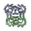
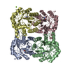
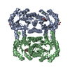
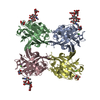
PDBj- Assembly
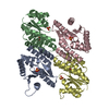
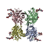
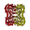
| Deposited unit | 
| ||||||||||||||||
|---|---|---|---|---|---|---|---|---|---|---|---|---|---|---|---|---|---|
| 1 |
| ||||||||||||||||
| Unit cell |
| ||||||||||||||||
| Noncrystallographic symmetry (NCS) | NCS oper:
|
-Components
| #1: Protein | Mass: 31087.818 Da / Num. of mol.: 4 / Fragment: RESIDUES 2-276 Source method: isolated from a genetically manipulated source Source: (gene. exp.) CANDIDATUS CLOACAMONAS ACIDAMINOVORANS (bacteria)Plasmid: PCRT7/CT-TOPO / Production host: #2: Chemical | ChemComp-ZN /   Mass: 65.409 Da / Num. of mol.: 4 / Source method: obtained synthetically / Formula: Zn Mass: 65.409 Da / Num. of mol.: 4 / Source method: obtained synthetically / Formula: Zn#3: Chemical |   Mass: 92.094 Da / Num. of mol.: 2 / Source method: obtained synthetically / Formula: C3H8O3 Mass: 92.094 Da / Num. of mol.: 2 / Source method: obtained synthetically / Formula: C3H8O3#4: Water | ChemComp-HOH / |  Mass: 18.015 Da / Num. of mol.: 976 / Source method: isolated from a natural source / Formula: H2O Mass: 18.015 Da / Num. of mol.: 976 / Source method: isolated from a natural source / Formula: H2O |
|---|
-Experimental details
-Experiment
| Experiment | Method: X-RAY DIFFRACTION / Number of used crystals: 1 |
|---|
- Sample preparation
| Crystal | Density Matthews: 2.24 Å3/Da / Density % sol: 45.22 % / Description: NONE |
|---|---|
| Crystal grow | pH: 7.5 Details: 30% PEG8000, 0.08 M NA ACETATE, 100 MM NA-HEPES PH 7.5 . |
-Data collection
| Diffraction | Mean temperature: 110 K |
|---|---|
| Diffraction source | Source: SYNCHROTRON / Site: SOLEIL  / Beamline: PROXIMA 1 / Wavelength: 0.9724 / Beamline: PROXIMA 1 / Wavelength: 0.9724 |
| Detector | Type: ADSC CCD / Detector: CCD / Date: Mar 27, 2008 / Details: KIRKPATRICK-BAEZ PAIR OF BI-MORPH MIRRORS |
| Radiation | Monochromator: CHANNEL CUT MONOCHROMATOR CRYSTAL / Protocol: SINGLE WAVELENGTH / Monochromatic (M) / Laue (L): M / Scattering type: x-ray |
| Radiation wavelength | Wavelength: 0.9724 Å / Relative weight: 1 |
| Reflection | Resolution: 1.59→19.47 Å / Num. obs: 147835 / % possible obs: 99.3 % / Observed criterion σ(I): 3 / Redundancy: 6.7 % / Biso Wilson estimate: 20.28 Å2 / Rmerge(I) obs: 0.07 / Net I/σ(I): 16.5 |
| Reflection shell | Resolution: 1.59→1.68 Å / Redundancy: 4.5 % / Rmerge(I) obs: 0.5 / Mean I/σ(I) obs: 3 / % possible all: 95.7 |
- Processing
| Software |
| |||||||||||||||||||||||||||||||||||||||||||||||||||||||||||||||||||||||||||||||||||||||||||||||||||||||||||||||||||||||||||||
|---|---|---|---|---|---|---|---|---|---|---|---|---|---|---|---|---|---|---|---|---|---|---|---|---|---|---|---|---|---|---|---|---|---|---|---|---|---|---|---|---|---|---|---|---|---|---|---|---|---|---|---|---|---|---|---|---|---|---|---|---|---|---|---|---|---|---|---|---|---|---|---|---|---|---|---|---|---|---|---|---|---|---|---|---|---|---|---|---|---|---|---|---|---|---|---|---|---|---|---|---|---|---|---|---|---|---|---|---|---|---|---|---|---|---|---|---|---|---|---|---|---|---|---|---|---|---|
| Refinement | Method to determine structure: MOLECULAR REPLACEMENT Starting model: PARTIAL MODEL FROM SEMET MAD PHASING IN DIFFERENT SPACE GROUP Resolution: 1.59→19.45 Å / Cor.coef. Fo:Fc: 0.9652 / Cor.coef. Fo:Fc free: 0.9586 / SU R Cruickshank DPI: 0.074 / Cross valid method: THROUGHOUT / σ(F): 0 / SU R Blow DPI: 0.076 / SU Rfree Blow DPI: 0.072 / SU Rfree Cruickshank DPI: 0.07 Details: IDEAL-DIST CONTACT TERM CONTACT SETUP. RESIDUE TYPES WITHOUT CCP4 ATOM TYPE IN LIBRARY=ZN GOL. NUMBER OF ATOMS WITH PROPER CCP4 ATOM TYPE=9408. NUMBER WITH APPROX DEFAULT CCP4 ATOM TYPE=12. ...Details: IDEAL-DIST CONTACT TERM CONTACT SETUP. RESIDUE TYPES WITHOUT CCP4 ATOM TYPE IN LIBRARY=ZN GOL. NUMBER OF ATOMS WITH PROPER CCP4 ATOM TYPE=9408. NUMBER WITH APPROX DEFAULT CCP4 ATOM TYPE=12. NUMBER TREATED BY BAD NON-BONDED CONTACTS=4.
| |||||||||||||||||||||||||||||||||||||||||||||||||||||||||||||||||||||||||||||||||||||||||||||||||||||||||||||||||||||||||||||
| Displacement parameters | Biso mean: 22.8 Å2
| |||||||||||||||||||||||||||||||||||||||||||||||||||||||||||||||||||||||||||||||||||||||||||||||||||||||||||||||||||||||||||||
| Refine analyze | Luzzati coordinate error obs: 0.154 Å | |||||||||||||||||||||||||||||||||||||||||||||||||||||||||||||||||||||||||||||||||||||||||||||||||||||||||||||||||||||||||||||
| Refinement step | Cycle: LAST / Resolution: 1.59→19.45 Å
| |||||||||||||||||||||||||||||||||||||||||||||||||||||||||||||||||||||||||||||||||||||||||||||||||||||||||||||||||||||||||||||
| Refine LS restraints |
| |||||||||||||||||||||||||||||||||||||||||||||||||||||||||||||||||||||||||||||||||||||||||||||||||||||||||||||||||||||||||||||
| LS refinement shell | Resolution: 1.59→1.63 Å / Total num. of bins used: 20
| |||||||||||||||||||||||||||||||||||||||||||||||||||||||||||||||||||||||||||||||||||||||||||||||||||||||||||||||||||||||||||||
| Refinement TLS params. | Method: refined / Refine-ID: X-RAY DIFFRACTION
| |||||||||||||||||||||||||||||||||||||||||||||||||||||||||||||||||||||||||||||||||||||||||||||||||||||||||||||||||||||||||||||
| Refinement TLS group |
|
