 Movie
Movie Controller

[English] 日本語
 Yorodumi
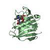
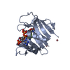
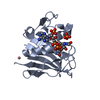
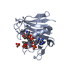
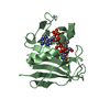
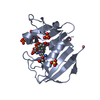
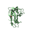
- PDB-2y6j: X-2 engineered mutated CBM4-2 Carbohydrate Binding Module from a ... -
+ Open data

- Basic information
| Entry | Database: PDB / ID: 2y6j | ||||||
|---|---|---|---|---|---|---|---|
| Title | X-2 engineered mutated CBM4-2 Carbohydrate Binding Module from a Thermostable Rhodothermus marinus Xylanase | ||||||
 Components Components | XYLANASE | ||||||
 Keywords Keywords | HYDROLASE | ||||||
| Function / homology |  Function and homology information Function and homology informationhydrolase activity, acting on glycosyl bonds / endo-1,4-beta-xylanase / endo-1,4-beta-xylanase activity / xylan catabolic process / metal ion binding Similarity search - Function | ||||||
| Biological species |   RHODOTHERMUS MARINUS (bacteria) RHODOTHERMUS MARINUS (bacteria) | ||||||
| Method |  X-RAY DIFFRACTION / SYNCHROTRON / MOLECULAR REPLACEMENT / Resolution: 1.7 Å X-RAY DIFFRACTION / SYNCHROTRON / MOLECULAR REPLACEMENT / Resolution: 1.7 Å | ||||||
 Authors Authors | von Schantz, L. / Hakansson, M. / Logan, D.T. / Walse, B. / Osterlin, J. / Nordberg-Karlsson, E. / Ohlin, M. | ||||||
 Citation Citation | Journal: Glycobiology / Year: 2012 Title: Structural basis for carbohydrate-binding specificity--a comparative assessment of two engineered carbohydrate-binding modules. Authors: von Schantz, L. / Hakansson, M. / Logan, D.T. / Walse, B. / Osterlin, J. / Nordberg-Karlsson, E. / Ohlin, M. | ||||||
| History |
|
- Structure visualization
| Structure viewer | Molecule: MolmilJmol/JSmol |
|---|
- Downloads & links
-Download
| PDBx/mmCIF format | 2y6j.cif.gz | 51.9 KB | Display | PDBx/mmCIF format |
|---|---|---|---|---|
| PDB format | pdb2y6j.ent.gz | 35.1 KB | Display | PDB format |
| PDBx/mmJSON format | 2y6j.json.gz | Tree view | PDBx/mmJSON format | |
| Others |  Other downloads Other downloads |
-Validation report
| Arichive directory | https://data.pdbj.org/pub/pdb/validation_reports/y6/2y6jftp://data.pdbj.org/pub/pdb/validation_reports/y6/2y6j | HTTPS FTP |
|---|
-Related structure data
| Related structure data |  2y64C  2y6gC 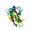 2y6hC  2y6kC  2y6lC  1k42S C: citing same article ( S: Starting model for refinement |
|---|---|
| Similar structure data |

-Links
 PDBj
PDBj
- Assembly
| Deposited unit | 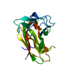
| ||||||||
|---|---|---|---|---|---|---|---|---|---|
| 1 |
| ||||||||
| Unit cell |
|
-Components
| #1: Protein | Mass: 17905.717 Da / Num. of mol.: 1 / Mutation: YES Source method: isolated from a genetically manipulated source Source: (gene. exp.) RHODOTHERMUS MARINUS (bacteria) / Production host: References: UniProt: Q6V8M0, UniProt: Q7WTN6*PLUS, endo-1,4-beta-xylanase |
|---|---|
| #2: Chemical | ChemComp-CA /   Mass: 40.078 Da / Num. of mol.: 1 / Source method: obtained synthetically / Formula: Ca Mass: 40.078 Da / Num. of mol.: 1 / Source method: obtained synthetically / Formula: Ca |
| #3: Water | ChemComp-HOH /  Mass: 18.015 Da / Num. of mol.: 201 / Source method: isolated from a natural source / Formula: H2O Mass: 18.015 Da / Num. of mol.: 201 / Source method: isolated from a natural source / Formula: H2O |
| Compound details | ENGINEERED RESIDUE IN CHAIN A, TRP 68 TO PHE ENGINEERED RESIDUE IN CHAIN A, ASP 69 TO ASN ...ENGINEERED |
| Sequence details | THIS CONSTRUCT STARTS WITH M (POSITION 1) AND ENDS WITH ONE L |
-Experimental details
-Experiment
| Experiment | Method: X-RAY DIFFRACTION / Number of used crystals: 1 |
|---|
- Sample preparation
| Crystal | Density Matthews: 2.1 Å3/Da / Density % sol: 41.2 % / Description: NONE |
|---|---|
| Crystal grow | pH: 6.5 / Details: 1.4 M SODIUM CITRATE PH 6.5 |
-Data collection
| Diffraction | Mean temperature: 100 K |
|---|---|
| Diffraction source | Source: SYNCHROTRON / Site: MAX II  / Beamline: I911-2 / Wavelength: 1.04 / Beamline: I911-2 / Wavelength: 1.04 |
| Detector | Type: MARRESEARCH SX-165 / Detector: CCD / Date: Dec 13, 2007 / Details: MIRRORS |
| Radiation | Monochromator: GERMANIUM CRYSTAL / Protocol: SINGLE WAVELENGTH / Monochromatic (M) / Laue (L): M / Scattering type: x-ray |
| Radiation wavelength | Wavelength: 1.04 Å / Relative weight: 1 |
| Reflection | Resolution: 1.7→17 Å / Num. obs: 16932 / % possible obs: 99.5 % / Observed criterion σ(I): -3 / Redundancy: 3.5 % / Rmerge(I) obs: 0.06 / Net I/σ(I): 15.6 |
| Reflection shell | Resolution: 1.7→1.74 Å / Rmerge(I) obs: 0.57 / Mean I/σ(I) obs: 2.3 / % possible all: 99.6 |
- Processing
| Software |
| ||||||||||||||||||||||||||||||||||||||||||||||||||||||||||||||||||||||||||||||||||||||||||||||||||||||||||||||||||||||||||||||||||||||||||||||||||||||||||||||||||||||||||||||||||||||
|---|---|---|---|---|---|---|---|---|---|---|---|---|---|---|---|---|---|---|---|---|---|---|---|---|---|---|---|---|---|---|---|---|---|---|---|---|---|---|---|---|---|---|---|---|---|---|---|---|---|---|---|---|---|---|---|---|---|---|---|---|---|---|---|---|---|---|---|---|---|---|---|---|---|---|---|---|---|---|---|---|---|---|---|---|---|---|---|---|---|---|---|---|---|---|---|---|---|---|---|---|---|---|---|---|---|---|---|---|---|---|---|---|---|---|---|---|---|---|---|---|---|---|---|---|---|---|---|---|---|---|---|---|---|---|---|---|---|---|---|---|---|---|---|---|---|---|---|---|---|---|---|---|---|---|---|---|---|---|---|---|---|---|---|---|---|---|---|---|---|---|---|---|---|---|---|---|---|---|---|---|---|---|---|
| Refinement | Method to determine structure: MOLECULAR REPLACEMENT Starting model: PDB ENTRY 1K42 Resolution: 1.7→17 Å / Cor.coef. Fo:Fc: 0.971 / Cor.coef. Fo:Fc free: 0.948 / SU B: 2.04 / SU ML: 0.066 / Cross valid method: THROUGHOUT / σ(F): 0 / ESU R: 0.102 / ESU R Free: 0.106 / Stereochemistry target values: MAXIMUM LIKELIHOOD / Details: HYDROGENS HAVE BEEN ADDED IN THE RIDING POSITIONS.
| ||||||||||||||||||||||||||||||||||||||||||||||||||||||||||||||||||||||||||||||||||||||||||||||||||||||||||||||||||||||||||||||||||||||||||||||||||||||||||||||||||||||||||||||||||||||
| Solvent computation | Ion probe radii: 0.8 Å / Shrinkage radii: 0.8 Å / VDW probe radii: 1.4 Å / Solvent model: BABINET MODEL WTH MASK | ||||||||||||||||||||||||||||||||||||||||||||||||||||||||||||||||||||||||||||||||||||||||||||||||||||||||||||||||||||||||||||||||||||||||||||||||||||||||||||||||||||||||||||||||||||||
| Displacement parameters | Biso mean: 14 Å2
| ||||||||||||||||||||||||||||||||||||||||||||||||||||||||||||||||||||||||||||||||||||||||||||||||||||||||||||||||||||||||||||||||||||||||||||||||||||||||||||||||||||||||||||||||||||||
| Refinement step | Cycle: LAST / Resolution: 1.7→17 Å
| ||||||||||||||||||||||||||||||||||||||||||||||||||||||||||||||||||||||||||||||||||||||||||||||||||||||||||||||||||||||||||||||||||||||||||||||||||||||||||||||||||||||||||||||||||||||
| Refine LS restraints |
|