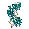
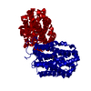
SHEET THE SHEET STRUCTURE OF THIS MOLECULE IS BIFURCATED. IN ORDER TO REPRESENT THIS FEATURE IN ... SHEET THE SHEET STRUCTURE OF THIS MOLECULE IS BIFURCATED. IN ORDER TO REPRESENT THIS FEATURE IN THE SHEET RECORDS BELOW, TWO SHEETS ARE DEFINED.
Mass: 18.015 Da / Num. of mol.: 473 / Source method: isolated from a natural source / Formula: H2O
-
Details
Nonpolymer details
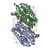
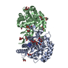
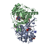
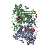
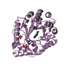
PYRIDOXAL-5'-PHOSPHATE (PLP) IS A PHYSIOLOGICAL CO-FACTOR.
Sequence details
THE PROTEIN BEARS AN N-TERMINAL HEXA-HISTIDINE PURIFICATION TAG NOT VISIBLE IN ELECTRON DENSITY.
-
Experimental details
-
Experiment
Experiment
Method: X-RAY DIFFRACTION / Number of used crystals: 1
-
Sample preparation
Crystal
Density Matthews: 2.31 Å3/Da / Density % sol: 42.42 % / Description: NONE
Crystal grow
Temperature: 293 K / Method: vapor diffusion, hanging drop / pH: 6.5 Details: VAPOUR DIFFUSION, HANGING DROP, 293K, 1 TO 1 MIXTURE OF RESERVOIR TO PROTEIN SOLUTION. RESERVOIR CONTAINED 1.8 M AMMONIUM SULFATE, 10 % W/V PEG400, 0.1 M MES PH 6.5. PROTEIN SOLUTION ...Details: VAPOUR DIFFUSION, HANGING DROP, 293K, 1 TO 1 MIXTURE OF RESERVOIR TO PROTEIN SOLUTION. RESERVOIR CONTAINED 1.8 M AMMONIUM SULFATE, 10 % W/V PEG400, 0.1 M MES PH 6.5. PROTEIN SOLUTION CONTAINED PABC PROTEIN AT 33 MG/ML, 500 MM NACL, 100 MM HEPES PH 7.5, 0.1 MM PYRIDOXAL PHOSPHATE AND 10 MM PARA-AMINOBENZOIC ACID.
Resolution: 1.75→101.34 Å / Cor.coef. Fo:Fc: 0.96 / Cor.coef. Fo:Fc free: 0.937 / SU B: 2.162 / SU ML: 0.07 / Cross valid method: THROUGHOUT / ESU R: 0.116 / ESU R Free: 0.118 / Stereochemistry target values: MAXIMUM LIKELIHOOD Details: HYDROGENS HAVE BEEN ADDED IN THE RIDING POSITIONS. U VALUES REFINED INDIVIDUALLY. THE SIDE-CHAIN FOR ARG 90 IN BOTH CHAIN A AND CHAIN B HAS ZERO OCCUPANCY BEYOND CB. THE PRESENCE OF THIS ...Details: HYDROGENS HAVE BEEN ADDED IN THE RIDING POSITIONS. U VALUES REFINED INDIVIDUALLY. THE SIDE-CHAIN FOR ARG 90 IN BOTH CHAIN A AND CHAIN B HAS ZERO OCCUPANCY BEYOND CB. THE PRESENCE OF THIS ARGININE IN THE PROTEIN SEQUENCE HAS BEEN CONFIRMED BY DNA SEQUENCING.
Rfactor
Num. reflection
% reflection
Selection details
Rfree
0.21543
2851
5.1 %
RANDOM
Rwork
0.16695
-
-
-
obs
0.16937
53372
98.42 %
-
Solvent computation
Ion probe radii: 0.8 Å / Shrinkage radii: 0.8 Å / VDW probe radii: 1.4 Å / Solvent model: MASK
 Movie
Movie Controller

 Yorodumi
 Open data
 Basic information
 Components
 Keywords
 Function and homology information
 PSEUDOMONAS AERUGINOSA (bacteria)
 X-RAY DIFFRACTION /
  Authors
 Citation
 Structure visualization
 Downloads & links
 Other downloads


 PDBj
PDBj
 Assembly
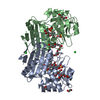


 Mass: 247.142 Da / Num. of mol.: 2 / Source method: obtained synthetically / Formula: C8H10NO6P
 Mass: 194.226 Da / Num. of mol.: 5 / Source method: obtained synthetically / Formula: C8H18O5 / Comment: precipitant*YM
 Mass: 35.453 Da / Num. of mol.: 3 / Source method: obtained synthetically / Formula: Cl
 Mass: 96.063 Da / Num. of mol.: 2 / Source method: obtained synthetically / Formula: SO4
 Mass: 62.068 Da / Num. of mol.: 3 / Source method: obtained synthetically / Formula: C2H6O2
 Mass: 106.120 Da / Num. of mol.: 3 / Source method: obtained synthetically / Formula: C4H10O3
 Mass: 92.094 Da / Num. of mol.: 2 / Source method: obtained synthetically / Formula: C3H8O3
 Sample preparation
 / Beamline: ID29 / Wavelength: 0.977
  Processing