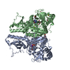
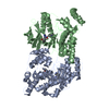
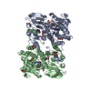
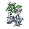
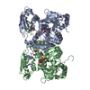
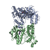
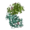
- PDB-2y27: crystal structure of PaaK1 in complex with ATP from Burkholderia ... -
+
Open data
ID or keywords:
Loading...
-
Basic information
Entry
Database: PDB / ID: 2y27
Title
crystal structure of PaaK1 in complex with ATP from Burkholderia cenocepacia
Components
PHENYLACETATE-COENZYME A LIGASE
Keywords
LIGASE / PHENYLACETIC ACID DEGRADATION PATHWAY
Function / homology
Function and homology information
phenylacetate-CoA ligase / phenylacetate-CoA ligase activity / phenylacetate catabolic process / ATP binding / metal ion binding Similarity search - Function
Journal: J.Biol.Chem. / Year: 2011 Title: Defining a Structural and Kinetic Rationale for Paralogous Copies of Phenylacetate-Coa Ligases from the Cystic Fibrosis Pathogen Burkholderia Cenocepacia J2315. Authors: Law, A. / Boulanger, M.J.
Protocol: SINGLE WAVELENGTH / Monochromatic (M) / Laue (L): M / Scattering type: x-ray
Radiation wavelength
Wavelength: 0.9792 Å / Relative weight: 1
Reflection
Resolution: 1.6→40 Å / Num. obs: 121839 / % possible obs: 95.8 % / Observed criterion σ(I): 2 / Redundancy: 4 % / Rmerge(I) obs: 0.08 / Net I/σ(I): 8.6
Reflection shell
Resolution: 1.6→1.69 Å / Redundancy: 4 % / Rmerge(I) obs: 0.5 / Mean I/σ(I) obs: 2.2 / % possible all: 94.1
-
Processing
Software
Name
Version
Classification
REFMAC
5.5.0109
refinement
iMOSFLM
datareduction
SCALA
datascaling
SHELXCDE
phasing
Refinement
Method to determine structure: SAD Starting model: NONE Resolution: 1.6→29.98 Å / Cor.coef. Fo:Fc: 0.972 / Cor.coef. Fo:Fc free: 0.961 / SU B: 1.705 / SU ML: 0.058 / Cross valid method: THROUGHOUT / ESU R: 0.082 / ESU R Free: 0.083 / Stereochemistry target values: MAXIMUM LIKELIHOOD / Details: HYDROGENS HAVE BEEN ADDED IN THE RIDING POSITIONS.
Rfactor
Num. reflection
% reflection
Selection details
Rfree
0.19407
6103
5 %
RANDOM
Rwork
0.16426
-
-
-
obs
0.16577
115707
95.76 %
-
Solvent computation
Ion probe radii: 0.8 Å / Shrinkage radii: 0.8 Å / VDW probe radii: 1.4 Å / Solvent model: MASK
 Movie
Movie Controller

 Yorodumi
 Open data
 Basic information
 Components
 Keywords
 Function and homology information
 BURKHOLDERIA CENOCEPACIA (bacteria)
 X-RAY DIFFRACTION /
  Authors
 Citation
 Structure visualization
 Downloads & links
 Other downloads
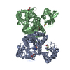


 PDBj
PDBj


 Assembly
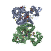


 Mass: 58.082 Da / Num. of mol.: 9 / Source method: obtained synthetically / Formula: CNS
 Mass: 92.094 Da / Num. of mol.: 5 / Source method: obtained synthetically / Formula: C3H8O3
 Mass: 78.133 Da / Num. of mol.: 5 / Source method: obtained synthetically / Formula: C2H6OS
 Mass: 194.226 Da / Num. of mol.: 1 / Source method: obtained synthetically / Formula: C8H18O5 / Comment: precipitant*YM
 Mass: 507.181 Da / Num. of mol.: 2 / Source method: obtained synthetically / Formula: C10H16N5O13P3 / Comment: ATP, energy-carrying molecule*YM
 Mass: 24.305 Da / Num. of mol.: 6 / Source method: obtained synthetically / Formula: Mg
 Sample preparation
 / Beamline: BL9-2 / Wavelength: 0.9792
  Processing