Monochromator: CU FILTER / Protocol: SINGLE WAVELENGTH / Monochromatic (M) / Laue (L): M / Scattering type: x-ray
Radiation wavelength
Wavelength: 0.9788 Å / Relative weight: 1
Reflection
Resolution: 2.4→33 Å / Num. obs: 5783 / % possible obs: 97.1 % / Observed criterion σ(I): 2.4 / Redundancy: 9.9 % / Biso Wilson estimate: 49.107 Å2 / Rmerge(I) obs: 0.06 / Net I/σ(I): 19.9
Reflection shell
Resolution: 2.4→2.5 Å / Redundancy: 4.4 % / Rmerge(I) obs: 0.25 / Mean I/σ(I) obs: 3.9 / % possible all: 81.2
-
Processing
Software
Name
Version
Classification
REFMAC
5.5.0102
refinement
MOSFLM
datareduction
SCALEPACK
datascaling
SHELXD
phasing
Refinement
Method to determine structure: SAD Starting model: NONE Resolution: 2.4→33 Å / Cor.coef. Fo:Fc: 0.94 / Cor.coef. Fo:Fc free: 0.934 / SU B: 20.845 / SU ML: 0.216 / Cross valid method: THROUGHOUT / ESU R: 0.431 / ESU R Free: 0.292 / Stereochemistry target values: MAXIMUM LIKELIHOOD / Details: HYDROGENS HAVE BEEN ADDED IN THE RIDING POSITIONS.
Rfactor
Num. reflection
% reflection
Selection details
Rfree
0.28412
627
9.8 %
RANDOM
Rwork
0.24093
-
-
-
obs
0.24523
5783
97.09 %
-
Solvent computation
Ion probe radii: 0.8 Å / Shrinkage radii: 0.8 Å / VDW probe radii: 1.4 Å / Solvent model: MASK
 Movie
Movie Controller


 Open data
 Basic information
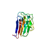
 Components
 Keywords
 Function and homology information
 HOMO SAPIENS (human)
 X-RAY DIFFRACTION /
  Authors
 Citation
 Structure visualization
 Downloads & links
 Other downloads


 PDBj
PDBj


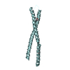
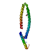
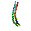
 Assembly

 Mass: 18.015 Da / Num. of mol.: 7 / Source method: isolated from a natural source / Formula: H2O
 Sample preparation
 / Beamline: X10SA / Wavelength: 0.9788
  Processing