| Entry | Database: PDB / ID: 2xtd
|
|---|
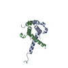
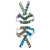
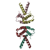
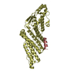
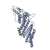
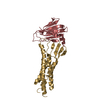
| Title | Structure of the TBL1 tetramerisation domain |
|---|
 Components Components | TBL1 F-BOX-LIKE/WD REPEAT-CONTAINING PROTEIN TBL1X |
|---|
 Keywords Keywords | TRANSCRIPTION / N-COR REPRESSOR COMPLEX / PROTEASOME |
|---|
| Function / homology |  Function and homology information Function and homology information
Loss of MECP2 binding ability to the NCoR/SMRT complex / Notch-HLH transcription pathway / histone deacetylase complex / Regulation of MECP2 expression and activity / NR1H3 & NR1H2 regulate gene expression linked to cholesterol transport and efflux / Regulation of lipid metabolism by PPARalpha / transcription repressor complex / BMAL1:CLOCK,NPAS2 activates circadian expression / RORA,B,C and NR1D1 (REV-ERBA) regulate gene expression / Activation of gene expression by SREBF (SREBP) ...Loss of MECP2 binding ability to the NCoR/SMRT complex / Notch-HLH transcription pathway / histone deacetylase complex / Regulation of MECP2 expression and activity / NR1H3 & NR1H2 regulate gene expression linked to cholesterol transport and efflux / Regulation of lipid metabolism by PPARalpha / transcription repressor complex / BMAL1:CLOCK,NPAS2 activates circadian expression / RORA,B,C and NR1D1 (REV-ERBA) regulate gene expression / Activation of gene expression by SREBF (SREBP) / Expression of BMAL (ARNTL), CLOCK, and NPAS2 / HDACs deacetylate histones / Heme signaling / PPARA activates gene expression / Transcriptional activation of mitochondrial biogenesis / Cytoprotection by HMOX1 / sensory perception of sound / Transcriptional regulation of white adipocyte differentiation / NOTCH1 Intracellular Domain Regulates Transcription / Constitutive Signaling by NOTCH1 PEST Domain Mutants / Constitutive Signaling by NOTCH1 HD+PEST Domain Mutants / HCMV Early Events / mitotic spindle / transcription corepressor activity / positive regulation of canonical Wnt signaling pathway / MLL4 and MLL3 complexes regulate expression of PPARG target genes in adipogenesis and hepatic steatosis / histone binding / proteasome-mediated ubiquitin-dependent protein catabolic process / transcription cis-regulatory region binding / protein stabilization / regulation of transcription by RNA polymerase II / positive regulation of DNA-templated transcription / DNA-templated transcription / negative regulation of transcription by RNA polymerase II / positive regulation of transcription by RNA polymerase II / proteolysis / nucleoplasm / identical protein binding / nucleusSimilarity search - Function Mitochondrial Import Receptor Subunit Tom20; Chain A - #30 / F-box-like/WD repeat-containing protein Ebi-like / LisH / Mitochondrial Import Receptor Subunit Tom20; Chain A / Lissencephaly type-1-like homology motif / LIS1 homology (LisH) motif profile. / LIS1 homology motif / Quinoprotein alcohol dehydrogenase-like superfamily / WD domain, G-beta repeat / G-protein beta WD-40 repeat ...Mitochondrial Import Receptor Subunit Tom20; Chain A - #30 / F-box-like/WD repeat-containing protein Ebi-like / LisH / Mitochondrial Import Receptor Subunit Tom20; Chain A / Lissencephaly type-1-like homology motif / LIS1 homology (LisH) motif profile. / LIS1 homology motif / Quinoprotein alcohol dehydrogenase-like superfamily / WD domain, G-beta repeat / G-protein beta WD-40 repeat / WD40 repeat, conserved site / Trp-Asp (WD) repeats signature. / Trp-Asp (WD) repeats profile. / Trp-Asp (WD) repeats circular profile. / WD40 repeats / WD40 repeat / WD40-repeat-containing domain superfamily / WD40/YVTN repeat-like-containing domain superfamily / Up-down Bundle / Mainly AlphaSimilarity search - Domain/homology |
|---|
| Biological species |  HOMO SAPIENS (human) HOMO SAPIENS (human) |
|---|
| Method |  X-RAY DIFFRACTION / SYNCHROTRON / MOLECULAR REPLACEMENT / Resolution: 3.2 Å X-RAY DIFFRACTION / SYNCHROTRON / MOLECULAR REPLACEMENT / Resolution: 3.2 Å |
|---|
 Authors Authors | Oberoi, J. / Fairall, L. / Watson, P.J. / Greenwood, J.A. / Schwabe, J.W.R. |
|---|
 Citation Citation | Journal: Nat.Struct.Mol.Biol. / Year: 2011
Title: Structural Basis for the Assembly of the Smrt/Ncor Core Transcriptional Repression Machinery.
Authors: Oberoi, J. / Fairall, L. / Watson, P.J. / Yang, J.C. / Czimmerer, Z. / Kampmann, T. / Goult, B.T. / Greenwood, J.A. / Gooch, J.T. / Kallenberger, B.C. / Nagy, L. / Neuhaus, D. / Schwabe, J.W.R. |
|---|
| History | | Deposition | Oct 6, 2010 | Deposition site: PDBE / Processing site: PDBE |
|---|
| Revision 1.0 | Jan 19, 2011 | Provider: repository / Type: Initial release |
|---|
| Revision 1.1 | May 8, 2011 | Group: Version format compliance |
|---|
| Revision 1.2 | Jul 13, 2011 | Group: Version format compliance |
|---|
| Revision 1.3 | Dec 20, 2023 | Group: Data collection / Database references ...Data collection / Database references / Other / Refinement description
Category: chem_comp_atom / chem_comp_bond ...chem_comp_atom / chem_comp_bond / database_2 / pdbx_database_status / pdbx_initial_refinement_model
Item: _database_2.pdbx_DOI / _database_2.pdbx_database_accession / _pdbx_database_status.status_code_sf |
|---|
|
|---|
 Movie
Movie Controller


 Open data
 Basic information
 Structure visualization
 Downloads & links
 Other downloads


 PDBj
PDBj


 Assembly


 Sample preparation
 / Beamline: ID14-2 / Wavelength: 0.934
  Processing