 Movie
Movie Controller


+ Open data

- Basic information
| Entry | Database: PDB / ID: 2xt0 | ||||||
|---|---|---|---|---|---|---|---|
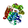
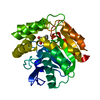
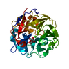
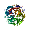
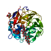
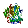
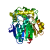
| Title | Dehalogenase DPpA from Plesiocystis pacifica SIR-I | ||||||
 Components Components | HALOALKANE DEHALOGENASE | ||||||
 Keywords Keywords | HYDROLASE / ALPHA-BETA HYDROLASE FOLD | ||||||
| Function / homology |  Function and homology information Function and homology information | ||||||
| Biological species |  PLESIOCYSTIS PACIFICA (bacteria) PLESIOCYSTIS PACIFICA (bacteria) | ||||||
| Method |  X-RAY DIFFRACTION / MOLECULAR REPLACEMENT / Resolution: 1.9 Å X-RAY DIFFRACTION / MOLECULAR REPLACEMENT / Resolution: 1.9 Å | ||||||
 Authors Authors | Bogdanovic, X. / Palm, G.J. / Hinrichs, W. | ||||||
 Citation Citation | Journal: Appl.Microbiol.Biotechnol. / Year: 2011 Title: Cloning, Functional Expression, Biochemical Characterization, and Structural Analysis of a Haloalkane Dehalogenase from Plesiocystis Pacifica Sir-1. Authors: Hesseler, M. / Bogdanovic, X. / Hidalgo, A. / Berenguer, J. / Palm, G.J. / Hinrichs, W. / Bornscheuer, U.T. #1: Journal: Acta Crystallogr.,Sect.F / Year: 2010 Title: Crystallization and Preliminary X-Ray Diffraction Studies of the Putative Haloalkane Dehalogenase Dppa from Plesiocystis Pacifica Sir-I. Authors: Bogdanovic, X. / Hesseler, M. / Palm, G.J. / Bornscheuer, U.T. / Hinrichs, W. | ||||||
| History |
|
- Structure visualization
| Structure viewer | Molecule: MolmilJmol/JSmol |
|---|
- Downloads & links
-Download
| PDBx/mmCIF format | 2xt0.cif.gz | 138.7 KB | Display | PDBx/mmCIF format |
|---|---|---|---|---|
| PDB format | pdb2xt0.ent.gz | 109.4 KB | Display | PDB format |
| PDBx/mmJSON format | 2xt0.json.gz | Tree view | PDBx/mmJSON format | |
| Others |  Other downloads Other downloads |
-Validation report
| Arichive directory | https://data.pdbj.org/pub/pdb/validation_reports/xt/2xt0ftp://data.pdbj.org/pub/pdb/validation_reports/xt/2xt0 | HTTPS FTP |
|---|
-Related structure data
| Related structure data |  1edbS S: Starting model for refinement |
|---|---|
| Similar structure data |


-Links
 PDBj
PDBj


- Assembly
| Deposited unit | 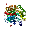
| ||||||||
|---|---|---|---|---|---|---|---|---|---|
| 1 |
| ||||||||
| 2 | 
| ||||||||
| Unit cell |
|
-Components
| #1: Protein | Mass: 32624.227 Da / Num. of mol.: 1 Source method: isolated from a genetically manipulated source Source: (gene. exp.) PLESIOCYSTIS PACIFICA (bacteria) / Strain: SIR-I / Production host: | ||
|---|---|---|---|
| #2: Chemical | ChemComp-SO4 /   Mass: 96.063 Da / Num. of mol.: 4 / Source method: obtained synthetically / Formula: SO4 Mass: 96.063 Da / Num. of mol.: 4 / Source method: obtained synthetically / Formula: SO4#3: Water | ChemComp-HOH / |  Mass: 18.015 Da / Num. of mol.: 260 / Source method: isolated from a natural source / Formula: H2O Mass: 18.015 Da / Num. of mol.: 260 / Source method: isolated from a natural source / Formula: H2O |
-Experimental details
-Experiment
| Experiment | Method: X-RAY DIFFRACTION / Number of used crystals: 1 |
|---|
- Sample preparation
| Crystal | Density Matthews: 2.6 Å3/Da / Density % sol: 53 % / Description: NONE |
|---|---|
| Crystal grow | pH: 6.5 / Details: 0.1 M MES PH 6.5, 1.8M (NH4)2SO4, 5% PEG400 |
-Data collection
| Diffraction | Mean temperature: 100 K |
|---|---|
| Diffraction source | Source: ROTATING ANODE / Type: RIGAKU MICROMAX-007 / Wavelength: 1.5418 |
| Detector | Type: RIGAKU SATURN 92 / Detector: CCD / Date: May 4, 2009 / Details: OSMIC MULTILAYER OPTIC |
| Radiation | Monochromator: OSMIC MULTILAYER OPTIC / Protocol: SINGLE WAVELENGTH / Monochromatic (M) / Laue (L): M / Scattering type: x-ray |
| Radiation wavelength | Wavelength: 1.5418 Å / Relative weight: 1 |
| Reflection | Resolution: 1.95→38.68 Å / Num. obs: 25613 / % possible obs: 98.5 % / Observed criterion σ(I): 0 / Redundancy: 5.5 % / Biso Wilson estimate: 26 Å2 / Rmerge(I) obs: 0.12 / Net I/σ(I): 7 |
| Reflection shell | Resolution: 1.95→2.02 Å / Redundancy: 4.1 % / Rmerge(I) obs: 0.44 / Mean I/σ(I) obs: 2.3 / % possible all: 97.4 |
- Processing
| Software |
| ||||||||||||||||||||||||||||||||||||||||||||||||||||||||||||||||||||||||||||||||||||||||||||||||||||||||||||||||||||||||||||||||||||||||||||||||||||||||||||||||||||||||||||||||||||||
|---|---|---|---|---|---|---|---|---|---|---|---|---|---|---|---|---|---|---|---|---|---|---|---|---|---|---|---|---|---|---|---|---|---|---|---|---|---|---|---|---|---|---|---|---|---|---|---|---|---|---|---|---|---|---|---|---|---|---|---|---|---|---|---|---|---|---|---|---|---|---|---|---|---|---|---|---|---|---|---|---|---|---|---|---|---|---|---|---|---|---|---|---|---|---|---|---|---|---|---|---|---|---|---|---|---|---|---|---|---|---|---|---|---|---|---|---|---|---|---|---|---|---|---|---|---|---|---|---|---|---|---|---|---|---|---|---|---|---|---|---|---|---|---|---|---|---|---|---|---|---|---|---|---|---|---|---|---|---|---|---|---|---|---|---|---|---|---|---|---|---|---|---|---|---|---|---|---|---|---|---|---|---|---|
| Refinement | Method to determine structure: MOLECULAR REPLACEMENT Starting model: PDB ENTRY 1EDB Resolution: 1.9→67.31 Å / Cor.coef. Fo:Fc: 0.943 / Cor.coef. Fo:Fc free: 0.93 / SU B: 8.419 / SU ML: 0.109 / Cross valid method: THROUGHOUT / ESU R: 0.176 / ESU R Free: 0.154 / Stereochemistry target values: MAXIMUM LIKELIHOOD Details: HYDROGENS HAVE BEEN ADDED IN THE RIDING POSITIONS. U VALUES WITH TLS ADDED. THE NUMBER OF UNIQUE REFLECTIONS FOR REFINEMENT IS 25826 AND FOR DATA PROCESSING 25613. THIS IS DUE TO A DIFFERENT ...Details: HYDROGENS HAVE BEEN ADDED IN THE RIDING POSITIONS. U VALUES WITH TLS ADDED. THE NUMBER OF UNIQUE REFLECTIONS FOR REFINEMENT IS 25826 AND FOR DATA PROCESSING 25613. THIS IS DUE TO A DIFFERENT RESOLUTION USED FOR REFINEMENT (1.9 A) AND PROCESSING (1.95 A).
| ||||||||||||||||||||||||||||||||||||||||||||||||||||||||||||||||||||||||||||||||||||||||||||||||||||||||||||||||||||||||||||||||||||||||||||||||||||||||||||||||||||||||||||||||||||||
| Solvent computation | Ion probe radii: 0.8 Å / Shrinkage radii: 0.8 Å / VDW probe radii: 1.4 Å / Solvent model: MASK | ||||||||||||||||||||||||||||||||||||||||||||||||||||||||||||||||||||||||||||||||||||||||||||||||||||||||||||||||||||||||||||||||||||||||||||||||||||||||||||||||||||||||||||||||||||||
| Displacement parameters | Biso mean: 20.519 Å2
| ||||||||||||||||||||||||||||||||||||||||||||||||||||||||||||||||||||||||||||||||||||||||||||||||||||||||||||||||||||||||||||||||||||||||||||||||||||||||||||||||||||||||||||||||||||||
| Refinement step | Cycle: LAST / Resolution: 1.9→67.31 Å
| ||||||||||||||||||||||||||||||||||||||||||||||||||||||||||||||||||||||||||||||||||||||||||||||||||||||||||||||||||||||||||||||||||||||||||||||||||||||||||||||||||||||||||||||||||||||
| Refine LS restraints |
|