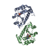
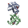
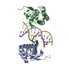
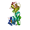
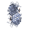
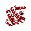
[English] 日本語
Yorodumi
- PDB-2xs6: CRYSTAL STRUCTURE OF THE RHOGAP DOMAIN OF HUMAN PIK3R2 -

+
Open data


ID or keywords:

Loading...

-
Basic information

Entry
Database: PDB / ID: 2xs6
TitleCRYSTAL STRUCTURE OF THE RHOGAP DOMAIN OF HUMAN PIK3R2
ComponentsPHOSPHATIDYLINOSITOL 3-KINASE REGULATORY SUBUNIT BETA
KeywordsTRANSFERASE / STRUCTURAL GENOMICS CONSORTIUM / SGC
Function / homology
Function and homology information


IRS-mediated signalling / regulation of actin filament polymerization / 1-phosphatidylinositol-3-kinase regulator activity / phosphatidylinositol 3-kinase regulatory subunit binding / RHOF GTPase cycle / regulation of stress fiber assembly / RHOD GTPase cycle / phosphatidylinositol 3-kinase complex, class IA / phosphatidylinositol 3-kinase complex / Nephrin family interactions ...IRS-mediated signalling / regulation of actin filament polymerization / 1-phosphatidylinositol-3-kinase regulator activity / phosphatidylinositol 3-kinase regulatory subunit binding / RHOF GTPase cycle / regulation of stress fiber assembly / RHOD GTPase cycle / phosphatidylinositol 3-kinase complex, class IA / phosphatidylinositol 3-kinase complex / Nephrin family interactions / RND1 GTPase cycle / Costimulation by the CD28 family / RND2 GTPase cycle / PI3K/AKT activation / RND3 GTPase cycle / negative regulation of MAPK cascade / Signaling by ALK / RHOB GTPase cycle / RHOJ GTPase cycle / intracellular glucose homeostasis / CD28 dependent PI3K/Akt signaling / Synthesis of PIPs at the plasma membrane / phosphatidylinositol phosphate biosynthetic process / CDC42 GTPase cycle / RHOU GTPase cycle / T cell differentiation / RET signaling / positive regulation of cell adhesion / Interleukin-3, Interleukin-5 and GM-CSF signaling / PI3K Cascade / RHOA GTPase cycle / RAC2 GTPase cycle / RAC3 GTPase cycle / regulation of protein localization to plasma membrane / Role of phospholipids in phagocytosis / Role of LAT2/NTAL/LAB on calcium mobilization / Interleukin receptor SHC signaling / Signaling by PDGFRA transmembrane, juxtamembrane and kinase domain mutants / Signaling by PDGFRA extracellular domain mutants / Tie2 Signaling / GPVI-mediated activation cascade / RAC1 GTPase cycle / Interleukin-7 signaling / phosphotyrosine residue binding / response to endoplasmic reticulum stress / Downstream signal transduction / B cell differentiation / phosphatidylinositol 3-kinase/protein kinase B signal transduction / Signaling by phosphorylated juxtamembrane, extracellular and kinase domain KIT mutants / regulation of autophagy / Regulation of signaling by CBL / Signaling by SCF-KIT / receptor tyrosine kinase binding / VEGFA-VEGFR2 Pathway / positive regulation of protein import into nucleus / cellular response to insulin stimulus / Constitutive Signaling by Aberrant PI3K in Cancer / Signaling by ALK fusions and activated point mutants / protein transport / Downstream TCR signaling / insulin receptor signaling pathway / PIP3 activates AKT signaling / DAP12 signaling / PI5P, PP2A and IER3 Regulate PI3K/AKT Signaling / RAF/MAP kinase cascade / G alpha (q) signalling events / protein phosphatase binding / Extra-nuclear estrogen signaling / immune response / protein heterodimerization activity / focal adhesion / positive regulation of transcription by RNA polymerase II / nucleus / cytosol
Similarity search - Function
Phosphatidylinositol 3-kinase regulatory subunit beta, SH3 domain / Phosphatidylinositol 3-kinase; Chain A / Rho GTPase activation protein / PI3K p85 subunit, C-terminal SH2 domain / PI3K regulatory subunit p85-related , inter-SH2 domain / PI3K p85 subunit, N-terminal SH2 domain / Phosphatidylinositol 3-kinase regulatory subunit P85 inter-SH2 domain / Rho GTPase-activating protein domain / RhoGAP domain / Rho GTPase-activating proteins domain profile. ...Phosphatidylinositol 3-kinase regulatory subunit beta, SH3 domain / Phosphatidylinositol 3-kinase; Chain A / Rho GTPase activation protein / PI3K p85 subunit, C-terminal SH2 domain / PI3K regulatory subunit p85-related , inter-SH2 domain / PI3K p85 subunit, N-terminal SH2 domain / Phosphatidylinositol 3-kinase regulatory subunit P85 inter-SH2 domain / Rho GTPase-activating protein domain / RhoGAP domain / Rho GTPase-activating proteins domain profile. / GTPase-activator protein for Rho-like GTPases / Rho GTPase activation protein / SH2 domain / Src homology 2 (SH2) domain profile. / Src homology 2 domains / SH2 domain / Src homology 3 domains / SH2 domain superfamily / SH3-like domain superfamily / Src homology 3 (SH3) domain profile. / SH3 domain / Orthogonal Bundle / Mainly Alpha
Similarity search - Domain/homology
Phosphatidylinositol 3-kinase regulatory subunit beta
Similarity search - Component
Biological speciesHOMO SAPIENS (human)
MethodX-RAY DIFFRACTION / SYNCHROTRON / MOLECULAR REPLACEMENT / Resolution: 2.09 Å
AuthorsTresaugues, L. / Welin, M. / Arrowsmith, C.H. / Berglund, H. / Bountra, C. / Collins, R. / Edwards, A.M. / Flodin, S. / Flores, A. / Graslund, S. ...Tresaugues, L. / Welin, M. / Arrowsmith, C.H. / Berglund, H. / Bountra, C. / Collins, R. / Edwards, A.M. / Flodin, S. / Flores, A. / Graslund, S. / Hammarstrom, M. / Johansson, I. / Karlberg, T. / Kol, S. / Kotenyova, T. / Kouznetsova, E. / Moche, M. / Nyman, T. / Persson, C. / Schuler, H. / Schutz, P. / Siponen, M.I. / Thorsell, A.G. / van der Berg, S. / Wahlberg, E. / Weigelt, J. / Nordlund, P.
CitationJournal: FEBS J. / Year: 2016
Title: The Structure and Catalytic Mechanism of Human Sphingomyelin Phosphodiesterase Like 3A - an Acid Sphingomyelinase Homolog with a Novel Nucleotide Hydrolase Activity.
Authors: Lim, S.M. / Yeung, K. / Tresaugues, L. / Ling, T.H. / Nordlund, P.
History
DepositionSep 24, 2010Deposition site: PDBE / Processing site: PDBE
Revision 1.0Nov 17, 2010Provider: repository / Type: Initial release
Revision 1.1Feb 3, 2016Group: Database references
Revision 1.2Apr 6, 2016Group: Database references
Revision 1.3Dec 20, 2023Group: Data collection / Database references ...Data collection / Database references / Derived calculations / Other / Refinement description
Category: chem_comp_atom / chem_comp_bond ...chem_comp_atom / chem_comp_bond / database_2 / pdbx_database_status / pdbx_initial_refinement_model / struct_site
Item: _database_2.pdbx_DOI / _database_2.pdbx_database_accession ..._database_2.pdbx_DOI / _database_2.pdbx_database_accession / _pdbx_database_status.status_code_sf / _struct_site.pdbx_auth_asym_id / _struct_site.pdbx_auth_comp_id / _struct_site.pdbx_auth_seq_id

-
Structure visualization

Structure viewerMolecule:
MolmilJmol/JSmol

Downloads & links

-
Assembly

Deposited unit
A: PHOSPHATIDYLINOSITOL 3-KINASE REGULATORY SUBUNIT BETA
hetero molecules


Theoretical massNumber of molelcules
Total (without water)22,9882
Polymers22,9531
Non-polymers351
Water1,45981
1
A: PHOSPHATIDYLINOSITOL 3-KINASE REGULATORY SUBUNIT BETA
hetero molecules

A: PHOSPHATIDYLINOSITOL 3-KINASE REGULATORY SUBUNIT BETA
hetero molecules


Theoretical massNumber of molelcules
Total (without water)45,9774
Polymers45,9062
Non-polymers712
Water362
TypeNameSymmetry operationNumber
identity operation1_555x,y,z1
crystal symmetry operation2_556-x,y,-z+11
Buried area1630 Å2
ΔGint-33 kcal/mol
Surface area15820 Å2
MethodPISA
Unit cell
Length a, b, c (Å)94.661, 34.354, 71.317
Angle α, β, γ (deg.)90.00, 130.88, 90.00
Int Tables number5
Space group name H-MC121
Components on special symmetry positions
IDModelComponents
11A-2025-

HOH

-
Components

#1: Protein PHOSPHATIDYLINOSITOL 3-KINASE REGULATORY SUBUNIT BETA / PHOSPHATIDYLINOSITOL 3-KINASE 85 KDA REGULATORY SUBUNIT BETA / PTDINS-3-KINASE REGULATORY SUBUNIT ...PHOSPHATIDYLINOSITOL 3-KINASE 85 KDA REGULATORY SUBUNIT BETA / PTDINS-3-KINASE REGULATORY SUBUNIT BETA / PI3-KINASE REGULATORY SUBUNIT BETA / PI3K REGULATORY SUBUNIT BETA / PTDINS-3-KINASE REGULATORY SUBUNIT P85-BETA / PI3-KINASE SUBUNIT P85-BETA


Mass: 22953.043 Da / Num. of mol.: 1 / Fragment: RHOGAP DOMAIN, RESIDUES 108-298
Source method: isolated from a genetically manipulated source
Source: (gene. exp.) HOMO SAPIENS (human) / Plasmid: PNIC-BSA4 / Production host: ESCHERICHIA COLI (E. coli) / Strain (production host): BL21(DE3)R3 PRARE / References: UniProt: O00459
#2: Chemical ChemComp-CL / CHLORIDE ION


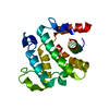
Mass: 35.453 Da / Num. of mol.: 1 / Source method: obtained synthetically / Formula: Cl
#3: Water ChemComp-HOH / water


Mass: 18.015 Da / Num. of mol.: 81 / Source method: isolated from a natural source / Formula: H2O

-
Experimental details

-
Experiment

ExperimentMethod: X-RAY DIFFRACTION / Number of used crystals: 1

-
Sample preparation

CrystalDensity Matthews: 1.91 Å3/Da / Density % sol: 35.7 % / Description: NONE
Crystal growpH: 6.5
Details: PROTEIN WAS CRYSTALLIZED FROM 0.1 M NACL, 0.85 M TRI-SODIUM CITRATE DIHYDRATE AND 0.1 M SODIUM CACODYLATE PH 6.5.

-
Data collection

DiffractionMean temperature: 100 K
Diffraction sourceSource: SYNCHROTRON / Site: BESSY / Beamline: 14.2 / Wavelength: 0.91841
DetectorType: MARRESEARCH / Detector: CCD / Date: Mar 17, 2010 / Details: MIRRORS
RadiationMonochromator: SI(111) / Protocol: SINGLE WAVELENGTH / Monochromatic (M) / Laue (L): M / Scattering type: x-ray
Radiation wavelengthWavelength: 0.91841 Å / Relative weight: 1
ReflectionResolution: 2.09→47.33 Å / Num. obs: 9968 / % possible obs: 95.6 % / Observed criterion σ(I): -3 / Redundancy: 3.7 % / Biso Wilson estimate: 29.6 Å2 / Rmerge(I) obs: 0.05 / Net I/σ(I): 9.9
Reflection shellResolution: 2.09→2.21 Å / Redundancy: 3.4 % / Rmerge(I) obs: 0.21 / Mean I/σ(I) obs: 3.7 / % possible all: 76.6

-
Processing

Software
NameVersionClassification
REFMAC5.5.0102refinement
XDSdata reduction
SCALAdata scaling
PHASERphasing
RefinementMethod to determine structure: MOLECULAR REPLACEMENT
Starting model: PDB ENTRY 1PBW
Resolution: 2.09→53.92 Å / Cor.coef. Fo:Fc: 0.949 / Cor.coef. Fo:Fc free: 0.903 / SU B: 10.993 / SU ML: 0.133 / Cross valid method: THROUGHOUT / ESU R: 0.243 / ESU R Free: 0.198 / Stereochemistry target values: MAXIMUM LIKELIHOOD / Details: HYDROGENS HAVE BEEN ADDED IN THE RIDING POSITIONS.
RfactorNum. reflection% reflectionSelection details
Rfree0.2398 563 5.6 %RANDOM
Rwork0.19149 ---
obs0.1942 9404 95.35 %-
Solvent computationIon probe radii: 0.8 Å / Shrinkage radii: 0.8 Å / VDW probe radii: 1.4 Å / Solvent model: MASK
Displacement parametersBiso mean: 29.775 Å2
Baniso -1Baniso -2Baniso -3
1--0.38 Å20 Å21.22 Å2
2--1.09 Å20 Å2
3---0.89 Å2
Refinement stepCycle: LAST / Resolution: 2.09→53.92 Å
ProteinNucleic acidLigandSolventTotal
Num. atoms1276 0 1 81 1358
Refine LS restraints
Refine-IDTypeDev idealDev ideal targetNumber
X-RAY DIFFRACTIONr_bond_refined_d0.0070.0221311
X-RAY DIFFRACTIONr_bond_other_d0.0010.02916
X-RAY DIFFRACTIONr_angle_refined_deg1.0322.0241799
X-RAY DIFFRACTIONr_angle_other_deg0.84732241
X-RAY DIFFRACTIONr_dihedral_angle_1_deg5.0165165
X-RAY DIFFRACTIONr_dihedral_angle_2_deg28.01522.54951
X-RAY DIFFRACTIONr_dihedral_angle_3_deg13.4815199
X-RAY DIFFRACTIONr_dihedral_angle_4_deg14.7221513
X-RAY DIFFRACTIONr_chiral_restr0.0490.2212
X-RAY DIFFRACTIONr_gen_planes_refined0.0050.0221425
X-RAY DIFFRACTIONr_gen_planes_other0.0010.02242
X-RAY DIFFRACTIONr_nbd_refined
X-RAY DIFFRACTIONr_nbd_other
X-RAY DIFFRACTIONr_nbtor_refined
X-RAY DIFFRACTIONr_nbtor_other
X-RAY DIFFRACTIONr_xyhbond_nbd_refined
X-RAY DIFFRACTIONr_xyhbond_nbd_other
X-RAY DIFFRACTIONr_metal_ion_refined
X-RAY DIFFRACTIONr_metal_ion_other
X-RAY DIFFRACTIONr_symmetry_vdw_refined
X-RAY DIFFRACTIONr_symmetry_vdw_other
X-RAY DIFFRACTIONr_symmetry_hbond_refined
X-RAY DIFFRACTIONr_symmetry_hbond_other
X-RAY DIFFRACTIONr_symmetry_metal_ion_refined
X-RAY DIFFRACTIONr_symmetry_metal_ion_other
X-RAY DIFFRACTIONr_mcbond_it0.3251.5846
X-RAY DIFFRACTIONr_mcbond_other0.0671.5316
X-RAY DIFFRACTIONr_mcangle_it0.60821368
X-RAY DIFFRACTIONr_mcangle_other
X-RAY DIFFRACTIONr_scbond_it0.9853465
X-RAY DIFFRACTIONr_scbond_other
X-RAY DIFFRACTIONr_scangle_it1.6464.5430
X-RAY DIFFRACTIONr_scangle_other
X-RAY DIFFRACTIONr_long_range_B_refined
X-RAY DIFFRACTIONr_long_range_B_other
X-RAY DIFFRACTIONr_rigid_bond_restr
X-RAY DIFFRACTIONr_sphericity_free
X-RAY DIFFRACTIONr_sphericity_bonded
LS refinement shellResolution: 2.094→2.148 Å / Total num. of bins used: 20
RfactorNum. reflection% reflection
Rfree0.245 26 -
Rwork0.24 461 -
obs--65.11 %
Refinement TLS params.

Method: refined / Refine-ID: X-RAY DIFFRACTION

IDL112)L122)L132)L222)L232)L332)S11 (Å °)S12 (Å °)S13 (Å °)S21 (Å °)S22 (Å °)S23 (Å °)S31 (Å °)S32 (Å °)S33 (Å °)T112)T122)T132)T222)T232)T332)Origin x (Å)Origin y (Å)Origin z (Å)
15.8262-0.0326-1.07294.7497-0.66843.9099-0.0644-0.0044-0.2163-0.118-0.0475-0.32170.02870.46740.11190.0955-0.02370.01220.0697-0.00320.1004-12.0329.794124.0523
27.4469-1.20524.52053.1979-3.159610.4285-0.24110.32560.146-0.1106-0.1203-0.1059-0.03440.16620.36150.07310.00420.03010.0204-0.02260.1058-13.58097.754319.0275
35.8123.858-15.11010.0084-3.873525.95380.56640.9798-0.1193-0.4264-0.07710.1316-1.7793-1.1173-0.48930.46850.1444-0.15060.45770.14570.8135-13.623617.6935-0.0837
416.1777-1.5597-0.29342.07550.68623.0201-0.11730.60170.913-0.09710.0253-0.073-0.27520.30.0920.1256-0.04090.00460.05490.06060.1059-6.737516.499415.0912
58.2242.59951.21338.4412-1.88358.35-0.23520.86710.1421-0.52770.17120.00970.08090.50250.0640.1459-0.00420.02860.21730.00040.0606-6.366710.69926.635
Refinement TLS group
IDRefine-IDRefine TLS-IDAuth asym-IDAuth seq-ID
1X-RAY DIFFRACTION1A109 - 160
2X-RAY DIFFRACTION2A161 - 194
3X-RAY DIFFRACTION3A195 - 212
4X-RAY DIFFRACTION4A213 - 242
5X-RAY DIFFRACTION5A243 - 294

+
About Yorodumi

-
News

-
Feb 9, 2022. New format data for meta-information of EMDB entries

New format data for meta-information of EMDB entries

  • Version 3 of the EMDB header file is now the official format.
  • The previous official version 1.9 will be removed from the archive.

Related info.:EMDB header

External links:wwPDB to switch to version 3 of the EMDB data model

-
Aug 12, 2020. Covid-19 info

Covid-19 info

URL: https://pdbjlvh1.pdbj.org/emnavi/covid19.php

New page: Covid-19 featured information page in EM Navigator.

Related info.:Covid-19 info / Mar 5, 2020. Novel coronavirus structure data

+
Mar 5, 2020. Novel coronavirus structure data

Novel coronavirus structure data

Related info.:Yorodumi Speices / Aug 12, 2020. Covid-19 info

External links:COVID-19 featured content - PDBj / Molecule of the Month (242):Coronavirus Proteases

+
Jan 31, 2019. EMDB accession codes are about to change! (news from PDBe EMDB page)

EMDB accession codes are about to change! (news from PDBe EMDB page)

  • The allocation of 4 digits for EMDB accession codes will soon come to an end. Whilst these codes will remain in use, new EMDB accession codes will include an additional digit and will expand incrementally as the available range of codes is exhausted. The current 4-digit format prefixed with “EMD-” (i.e. EMD-XXXX) will advance to a 5-digit format (i.e. EMD-XXXXX), and so on. It is currently estimated that the 4-digit codes will be depleted around Spring 2019, at which point the 5-digit format will come into force.
  • The EM Navigator/Yorodumi systems omit the EMD- prefix.

Related info.:Q: What is EMD? / ID/Accession-code notation in Yorodumi/EM Navigator

External links:EMDB Accession Codes are Changing Soon! / Contact to PDBj

+
Jul 12, 2017. Major update of PDB

Major update of PDB

  • wwPDB released updated PDB data conforming to the new PDBx/mmCIF dictionary.
  • This is a major update changing the version number from 4 to 5, and with Remediation, in which all the entries are updated.
  • In this update, many items about electron microscopy experimental information are reorganized (e.g. em_software).
  • Now, EM Navigator and Yorodumi are based on the updated data.

External links:wwPDB Remediation / Enriched Model Files Conforming to OneDep Data Standards Now Available in the PDB FTP Archive

-
Yorodumi

Thousand views of thousand structures

  • Yorodumi is a browser for structure data from EMDB, PDB, SASBDB, etc.
  • This page is also the successor to EM Navigator detail page, and also detail information page/front-end page for Omokage search.
  • The word "yorodu" (or yorozu) is an old Japanese word meaning "ten thousand". "mi" (miru) is to see.

Related info.:EMDB / PDB / SASBDB / Comparison of 3 databanks / Yorodumi Search / Aug 31, 2016. New EM Navigator & Yorodumi / Yorodumi Papers / Jmol/JSmol / Function and homology information / Changes in new EM Navigator and Yorodumi

Read more