Protocol: SINGLE WAVELENGTH / Monochromatic (M) / Laue (L): M / Scattering type: x-ray
Radiation wavelength
Wavelength: 0.88567 Å / Relative weight: 1
Reflection
Resolution: 1.5→19.94 Å / Num. obs: 95566 / % possible obs: 98.4 % / Observed criterion σ(I): -3 / Redundancy: 5.4 % / Biso Wilson estimate: 12.6 Å2 / Rmerge(I) obs: 0.08 / Net I/σ(I): 11.8
Reflection shell
Resolution: 1.5→1.58 Å / Redundancy: 5.4 % / Rmerge(I) obs: 0.39 / Mean I/σ(I) obs: 4.4 / % possible all: 98.8
-
Processing
Software
Name
Version
Classification
REFMAC
5.5.0109
refinement
XDS
datareduction
XSCALE
datascaling
PHASER
phasing
Refinement
Method to determine structure: MOLECULAR REPLACEMENT Starting model: UNPUBLISHED STRUCTURAL DATA OF M. BARKERI PHOTOLYASE Resolution: 1.5→15 Å / Cor.coef. Fo:Fc: 0.965 / Cor.coef. Fo:Fc free: 0.952 / SU B: 3.055 / SU ML: 0.049 / Cross valid method: THROUGHOUT / ESU R: 0.066 / ESU R Free: 0.068 / Stereochemistry target values: MAXIMUM LIKELIHOOD Details: HYDROGENS HAVE BEEN ADDED IN THE RIDING POSITIONS. U VALUES WITH TLS ADDED. RESIDUES 189-197 ARE DISORDERED.
Rfactor
Num. reflection
% reflection
Selection details
Rfree
0.21017
1416
1.5 %
RANDOM
Rwork
0.18486
-
-
-
obs
0.18523
94089
98.26 %
-
Solvent computation
Ion probe radii: 0.8 Å / Shrinkage radii: 0.8 Å / VDW probe radii: 1.2 Å / Solvent model: MASK
 Movie
Movie Controller

 Yorodumi
 Open data
 Basic information
 Components
 Keywords
 Function and homology information
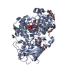
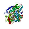
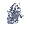
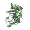
 METHANOSARCINA MAZEI (archaea)
 X-RAY DIFFRACTION /
  Authors
 Citation
 Structure visualization
 Downloads & links
 Other downloads


 PDBj
PDBj
 Assembly


 Mass: 785.550 Da / Num. of mol.: 1 / Source method: obtained synthetically / Formula: C27H33N9O15P2 / Comment: FAD*YM
 Mass: 96.063 Da / Num. of mol.: 9 / Source method: obtained synthetically / Formula: SO4
 Mass: 92.094 Da / Num. of mol.: 2 / Source method: obtained synthetically / Formula: C3H8O3
 Mass: 18.015 Da / Num. of mol.: 375 / Source method: isolated from a natural source / Formula: H2O
 Sample preparation
 / Beamline: ID29 / Wavelength: 0.88567
  Processing