 Movie
Movie Controller

[English] 日本語
 Yorodumi
- PDB-2xr0: Room temperature X-ray structure of the perdeuterated Toho-1 R274... -
+ Open data

- Basic information
| Entry | Database: PDB / ID: 2xr0 | ||||||
|---|---|---|---|---|---|---|---|
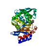
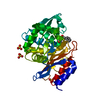
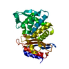
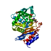
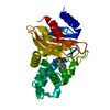
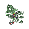
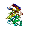
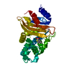
| Title | Room temperature X-ray structure of the perdeuterated Toho-1 R274N R276N double mutant beta-lactamase | ||||||
 Components Components | TOHO-1 BETA-LACTAMASE | ||||||
 Keywords Keywords | HYDROLASE / EXTENDED-SPECTRUM BETA-LACTAMASES / ESBLS / CTX-M-TYPE ESBLS | ||||||
| Function / homology |  Function and homology information Function and homology informationbeta-lactam antibiotic catabolic process / beta-lactamase activity / beta-lactamase / response to antibiotic Similarity search - Function | ||||||
| Biological species |  | ||||||
| Method |  X-RAY DIFFRACTION / SYNCHROTRON / MOLECULAR REPLACEMENT / Resolution: 2.2 Å X-RAY DIFFRACTION / SYNCHROTRON / MOLECULAR REPLACEMENT / Resolution: 2.2 Å | ||||||
 Authors Authors | Tomanicek, S.J. / Wang, K.K. / Weiss, K.L. / Blakeley, M.P. / Cooper, J. / Chen, Y. / Coates, L. | ||||||
 Citation Citation | Journal: FEBS Lett. / Year: 2011 Title: The Active Site Protonation States of Perdeuterated Toho-1 Beta-Lactamase Determined by Neutron Diffraction Support a Role for Glu166 as the General Base in Acylation. Authors: Tomanicek, S.J. / Wang, K.K. / Weiss, K.L. / Blakeley, M.P. / Cooper, J. / Chen, Y. / Coates, L. | ||||||
| History |
|
- Structure visualization
| Structure viewer | Molecule: MolmilJmol/JSmol |
|---|
- Downloads & links
-Download
| PDBx/mmCIF format | 2xr0.cif.gz | 66.8 KB | Display | PDBx/mmCIF format |
|---|---|---|---|---|
| PDB format | pdb2xr0.ent.gz | 48.4 KB | Display | PDB format |
| PDBx/mmJSON format | 2xr0.json.gz | Tree view | PDBx/mmJSON format | |
| Others |  Other downloads Other downloads |
-Validation report
| Arichive directory | https://data.pdbj.org/pub/pdb/validation_reports/xr/2xr0ftp://data.pdbj.org/pub/pdb/validation_reports/xr/2xr0 | HTTPS FTP |
|---|
-Related structure data
| Related structure data |  2xqzC  2wyxS S: Starting model for refinement C: citing same article ( |
|---|---|
| Similar structure data |


-Links
 PDBj
PDBj


- Assembly
| Deposited unit | 
| ||||||||
|---|---|---|---|---|---|---|---|---|---|
| 1 |
| ||||||||
| Unit cell |
|
-Components
| #1: Protein | Mass: 27990.607 Da / Num. of mol.: 1 / Fragment: RESIDUES 32-291 / Mutation: YES Source method: isolated from a genetically manipulated source Details: FULLY PERDEUTERATED / Source: (gene. exp.) | ||||
|---|---|---|---|---|---|
| #2: Chemical |   Mass: 96.063 Da / Num. of mol.: 3 / Source method: obtained synthetically / Formula: SO4 Mass: 96.063 Da / Num. of mol.: 3 / Source method: obtained synthetically / Formula: SO4#3: Water | ChemComp-HOH / |  Mass: 18.015 Da / Num. of mol.: 154 / Source method: isolated from a natural source / Formula: H2O Mass: 18.015 Da / Num. of mol.: 154 / Source method: isolated from a natural source / Formula: H2OSequence details | 2XR0 USES A RESIDUE NUMBERING SCHEME IN WHICH RESIDUE POSITION 58, RESIDUE POSITION 239, AND ...2XR0 USES A RESIDUE NUMBERING SCHEME IN WHICH RESIDUE POSITION 58, RESIDUE POSITION 239, AND RESIDUE POSITION 253 HAVE NOT BEEN USED - GIVING A SMALL MISMATCH BETWEEN THE NUMBER OF RESIDUES IN THE DBREF SEQUENCE RANGES BELOW. | |
-Experimental details
-Experiment
| Experiment | Method: X-RAY DIFFRACTION / Number of used crystals: 1 |
|---|
- Sample preparation
| Crystal | Density Matthews: 2.77 Å3/Da / Density % sol: 55.55 % / Description: NONE |
|---|---|
| Crystal grow | pH: 6.5 Details: 2.0 A AMMONIUM SULFATE, 0.1 M SODIUM CITRATE, PD 5.1. PREPARED IN 99.9% D2O AND MIXED 1:1 WITH PERDEUTERATED PROTEIN PREPARED IN 20 MM NA MES AT PD 6.1 (PH 6.5). |
-Data collection
| Diffraction | Mean temperature: 293 K |
|---|---|
| Diffraction source | Source: SYNCHROTRON / Site: APS  / Beamline: 22-ID / Wavelength: 1 / Beamline: 22-ID / Wavelength: 1 |
| Detector | Type: MARRESEARCH MX-300 / Detector: CCD / Date: Apr 4, 2010 |
| Radiation | Protocol: SINGLE WAVELENGTH / Monochromatic (M) / Laue (L): M / Scattering type: x-ray |
| Radiation wavelength | Wavelength: 1 Å / Relative weight: 1 |
| Reflection | Resolution: 2.2→29.54 Å / Num. obs: 15962 / % possible obs: 98.6 % / Observed criterion σ(I): 2 / Redundancy: 7.5 % / Biso Wilson estimate: 16.86 Å2 / Rmerge(I) obs: 0.05 / Net I/σ(I): 31.3 |
| Reflection shell | Resolution: 2.2→2.32 Å / Redundancy: 7.5 % / Rmerge(I) obs: 0.83 / Mean I/σ(I) obs: 22 / % possible all: 97.9 |
- Processing
| Software |
| |||||||||||||||||||||||||||||||||||||||||||||||||
|---|---|---|---|---|---|---|---|---|---|---|---|---|---|---|---|---|---|---|---|---|---|---|---|---|---|---|---|---|---|---|---|---|---|---|---|---|---|---|---|---|---|---|---|---|---|---|---|---|---|---|
| Refinement | Method to determine structure: MOLECULAR REPLACEMENT Starting model: PDB ENTRY 2WYX Resolution: 2.2→29.54 Å / SU ML: 0.16 / σ(F): 1.43 / Phase error: 16.06 / Stereochemistry target values: ML
| |||||||||||||||||||||||||||||||||||||||||||||||||
| Solvent computation | Shrinkage radii: 0.95 Å / VDW probe radii: 1.2 Å / Solvent model: FLAT BULK SOLVENT MODEL / Bsol: 28.96 Å2 / ksol: 0.33 e/Å3 | |||||||||||||||||||||||||||||||||||||||||||||||||
| Displacement parameters | Biso mean: 19.65 Å2
| |||||||||||||||||||||||||||||||||||||||||||||||||
| Refinement step | Cycle: LAST / Resolution: 2.2→29.54 Å
| |||||||||||||||||||||||||||||||||||||||||||||||||
| Refine LS restraints |
| |||||||||||||||||||||||||||||||||||||||||||||||||
| LS refinement shell |
|
