 Movie
Movie Controller

[English] 日本語
 Yorodumi
- PDB-2xoy: C-terminal cysteine-rich domain of human CHFR bound to P(1),P(2)-... -
+ Open data

- Basic information
| Entry | Database: PDB / ID: 2xoy | ||||||
|---|---|---|---|---|---|---|---|
| Title | C-terminal cysteine-rich domain of human CHFR bound to P(1),P(2)- Diadenosine-5'-pyrophosphate | ||||||
 Components Components | E3 UBIQUITIN-PROTEIN LIGASE CHFR | ||||||
 Keywords Keywords | LIGASE / ZINC-BINDING / PBZ / MITOSIS / ANTEPHASE CHECKPOINT | ||||||
| Function / homology |  Function and homology information Function and homology informationmeiotic spindle checkpoint signaling / mitotic G2/M transition checkpoint / positive regulation of protein ubiquitination / PML body / RING-type E3 ubiquitin transferase / protein destabilization / protein polyubiquitination / ubiquitin-protein transferase activity / ubiquitin protein ligase activity / positive regulation of proteasomal ubiquitin-dependent protein catabolic process ...meiotic spindle checkpoint signaling / mitotic G2/M transition checkpoint / positive regulation of protein ubiquitination / PML body / RING-type E3 ubiquitin transferase / protein destabilization / protein polyubiquitination / ubiquitin-protein transferase activity / ubiquitin protein ligase activity / positive regulation of proteasomal ubiquitin-dependent protein catabolic process / ubiquitin-dependent protein catabolic process / cell division / nucleotide binding / zinc ion binding / nucleus Similarity search - Function | ||||||
| Biological species |  HOMO SAPIENS (human) HOMO SAPIENS (human) | ||||||
| Method |  X-RAY DIFFRACTION / SYNCHROTRON / MOLECULAR REPLACEMENT / Resolution: 2.6 Å X-RAY DIFFRACTION / SYNCHROTRON / MOLECULAR REPLACEMENT / Resolution: 2.6 Å | ||||||
 Authors Authors | Oberoi, J. / Bayliss, R. | ||||||
 Citation Citation | Journal: J.Biol.Chem. / Year: 2010 Title: Structural Basis of Poly(Adp-Ribose) Recognition by the Multizinc Binding Domain of Checkpoint with Forkhead-Associated and Ring Domains (Chfr). Authors: Oberoi, J. / Richards, M.W. / Crumpler, S. / Brown, N. / Blagg, J. / Bayliss, R. | ||||||
| History |
|
- Structure visualization
| Structure viewer | Molecule: MolmilJmol/JSmol |
|---|
- Downloads & links
-Download
| PDBx/mmCIF format | 2xoy.cif.gz | 102.1 KB | Display | PDBx/mmCIF format |
|---|---|---|---|---|
| PDB format | pdb2xoy.ent.gz | 76.8 KB | Display | PDB format |
| PDBx/mmJSON format | 2xoy.json.gz | Tree view | PDBx/mmJSON format | |
| Others |  Other downloads Other downloads |
-Validation report
| Arichive directory | https://data.pdbj.org/pub/pdb/validation_reports/xo/2xoyftp://data.pdbj.org/pub/pdb/validation_reports/xo/2xoy | HTTPS FTP |
|---|
-Related structure data
| Related structure data |  2xocSC  2xozC  2xp0C S: Starting model for refinement C: citing same article ( |
|---|---|
| Similar structure data |


-Links
 PDBj
PDBj


- Assembly
| Deposited unit | 
| ||||||||
|---|---|---|---|---|---|---|---|---|---|
| 1 | 
| ||||||||
| 2 | 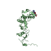
| ||||||||
| Unit cell |
|
-Components
| #1: Protein | Mass: 29268.252 Da / Num. of mol.: 2 / Fragment: CYSTEINE-RICH REGION, RESIDUES 407-664 Source method: isolated from a genetically manipulated source Source: (gene. exp.) HOMO SAPIENS (human) / Production host:  References: UniProt: Q96EP1, Ligases; Forming carbon-nitrogen bonds; Acid-amino-acid ligases (peptide synthases) #2: Chemical | ChemComp-ZN /   Mass: 65.409 Da / Num. of mol.: 10 / Source method: obtained synthetically / Formula: Zn Mass: 65.409 Da / Num. of mol.: 10 / Source method: obtained synthetically / Formula: Zn#3: Chemical | ChemComp-AMP / |   Mass: 347.221 Da / Num. of mol.: 1 / Source method: obtained synthetically / Formula: C10H14N5O7P / Comment: AMP*YM Mass: 347.221 Da / Num. of mol.: 1 / Source method: obtained synthetically / Formula: C10H14N5O7P / Comment: AMP*YM#4: Chemical | ChemComp-ADE / |   Mass: 135.127 Da / Num. of mol.: 1 / Source method: obtained synthetically / Formula: C5H5N5 Mass: 135.127 Da / Num. of mol.: 1 / Source method: obtained synthetically / Formula: C5H5N5#5: Water | ChemComp-HOH / |  Mass: 18.015 Da / Num. of mol.: 86 / Source method: isolated from a natural source / Formula: H2O Mass: 18.015 Da / Num. of mol.: 86 / Source method: isolated from a natural source / Formula: H2OHas protein modification | Y | Sequence details | N-TERMINAL SEQUENCE 'GAM' FROM VECTOR | |
|---|
-Experimental details
-Experiment
| Experiment | Method: X-RAY DIFFRACTION |
|---|
- Sample preparation
| Crystal | Density Matthews: 2.96 Å3/Da / Density % sol: 58.51 % / Description: NONE |
|---|---|
| Crystal grow | Details: 12% PEG 20000, 0.1M MES PH6.5, 0.1M KCL |
-Data collection
| Diffraction | Mean temperature: 100 K |
|---|---|
| Diffraction source | Source: SYNCHROTRON / Site: Diamond  / Beamline: I03 / Wavelength: 0.9763 / Beamline: I03 / Wavelength: 0.9763 |
| Detector | Type: ADSC CCD / Detector: CCD |
| Radiation | Protocol: SINGLE WAVELENGTH / Monochromatic (M) / Laue (L): M / Scattering type: x-ray |
| Radiation wavelength | Wavelength: 0.9763 Å / Relative weight: 1 |
| Reflection | Resolution: 2.6→65.63 Å / Num. obs: 21061 / % possible obs: 99.5 % / Observed criterion σ(I): 6 / Redundancy: 3.4 % / Biso Wilson estimate: 40.3 Å2 / Rmerge(I) obs: 0.09 / Net I/σ(I): 8.6 |
| Reflection shell | Resolution: 2.6→2.74 Å / Redundancy: 3.5 % / Rmerge(I) obs: 0.43 / Mean I/σ(I) obs: 2.6 / % possible all: 99.8 |
- Processing
| Software |
| |||||||||||||||||||||||||||||||||||||||||||||||||||||||||||||||||||||||||||||
|---|---|---|---|---|---|---|---|---|---|---|---|---|---|---|---|---|---|---|---|---|---|---|---|---|---|---|---|---|---|---|---|---|---|---|---|---|---|---|---|---|---|---|---|---|---|---|---|---|---|---|---|---|---|---|---|---|---|---|---|---|---|---|---|---|---|---|---|---|---|---|---|---|---|---|---|---|---|---|
| Refinement | Method to determine structure: MOLECULAR REPLACEMENT Starting model: PDB ENTRY 2XOC Resolution: 2.6→36.256 Å / SU ML: 0.36 / σ(F): 0.01 / Phase error: 24.37 / Stereochemistry target values: ML Details: P(1),P(2)-DIADENOSINE-5'- PYROPHOSPHATE MODELED AS AMP AND ADENINE.
| |||||||||||||||||||||||||||||||||||||||||||||||||||||||||||||||||||||||||||||
| Solvent computation | Shrinkage radii: 0.9 Å / VDW probe radii: 1.11 Å / Solvent model: FLAT BULK SOLVENT MODEL / Bsol: 21.63 Å2 / ksol: 0.299 e/Å3 | |||||||||||||||||||||||||||||||||||||||||||||||||||||||||||||||||||||||||||||
| Displacement parameters |
| |||||||||||||||||||||||||||||||||||||||||||||||||||||||||||||||||||||||||||||
| Refinement step | Cycle: LAST / Resolution: 2.6→36.256 Å
| |||||||||||||||||||||||||||||||||||||||||||||||||||||||||||||||||||||||||||||
| Refine LS restraints |
| |||||||||||||||||||||||||||||||||||||||||||||||||||||||||||||||||||||||||||||
| LS refinement shell |
|
