Mass: 18.015 Da / Num. of mol.: 416 / Source method: isolated from a natural source / Formula: H2O
-
Details
Compound details
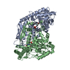
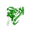
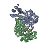
ENGINEERED RESIDUE IN CHAIN A, MET 1 TO GLU ENGINEERED RESIDUE IN CHAIN B, MET 1 TO GLU
Nonpolymer details
BF5-PLP: COVALENT LINK BETWEEN THE PLP COFACTOR AND THE SYNTHETIC INHIBITOR NAMED BFF-122.
Sequence details
THE SEGMENT 20-32 IS MISSING FROM THE COORDINATES FILE. THE FIRST RESIDUE IS A GLU THAT REPLACES A ...THE SEGMENT 20-32 IS MISSING FROM THE COORDINATES FILE. THE FIRST RESIDUE IS A GLU THAT REPLACES A MET AS A RESULT OF CLONING STRATEGY
-
Experimental details
-
Experiment
Experiment
Method: X-RAY DIFFRACTION / Number of used crystals: 1
-
Sample preparation
Crystal
Density Matthews: 2.41 Å3/Da / Density % sol: 48.96 % / Description: NONE
Resolution: 2.1→32.04 Å / Cor.coef. Fo:Fc: 0.941 / Cor.coef. Fo:Fc free: 0.902 / SU B: 4.583 / SU ML: 0.126 / Cross valid method: THROUGHOUT / ESU R: 0.209 / ESU R Free: 0.185 / Stereochemistry target values: MAXIMUM LIKELIHOOD / Details: HYDROGENS HAVE BEEN ADDED IN THE RIDING POSITIONS.
Rfactor
Num. reflection
% reflection
Selection details
Rfree
0.24123
1113
2.1 %
RANDOM
Rwork
0.18814
-
-
-
obs
0.18929
53172
100 %
-
Solvent computation
Ion probe radii: 0.8 Å / Shrinkage radii: 0.8 Å / VDW probe radii: 1.2 Å / Solvent model: BABINET MODEL WITH MASK
 Movie
Movie Controller


 Open data
 Basic information
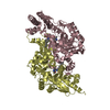
 Components
 Keywords
 Function and homology information
 HOMO SAPIENS (human)
 X-RAY DIFFRACTION /
  Authors
 Citation
 Structure visualization
 Downloads & links
 Other downloads


 PDBj
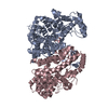
PDBj Assembly
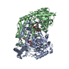

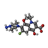


 Mass: 362.356 Da / Num. of mol.: 2 / Source method: obtained synthetically / Formula: C17H19FN4O4
 Mass: 247.142 Da / Num. of mol.: 2 / Source method: obtained synthetically / Formula: C8H10NO6P
 Mass: 126.904 Da / Num. of mol.: 1 / Source method: obtained synthetically / Formula: I
 Mass: 92.094 Da / Num. of mol.: 1 / Source method: obtained synthetically / Formula: C3H8O3
 Sample preparation
 / Beamline: ID14-1 / Wavelength: 0.981
  Processing